Review
. 2017 Nov 8;6(11):e96.
doi: 10.1038/emi.2017.82.

Vector competence of European mosquitoes for West Nile virus

Affiliations
Review

Vector competence of European mosquitoes for West Nile virus

Chantal Bf Vogels et al. Emerg Microbes Infect. .

Abstract

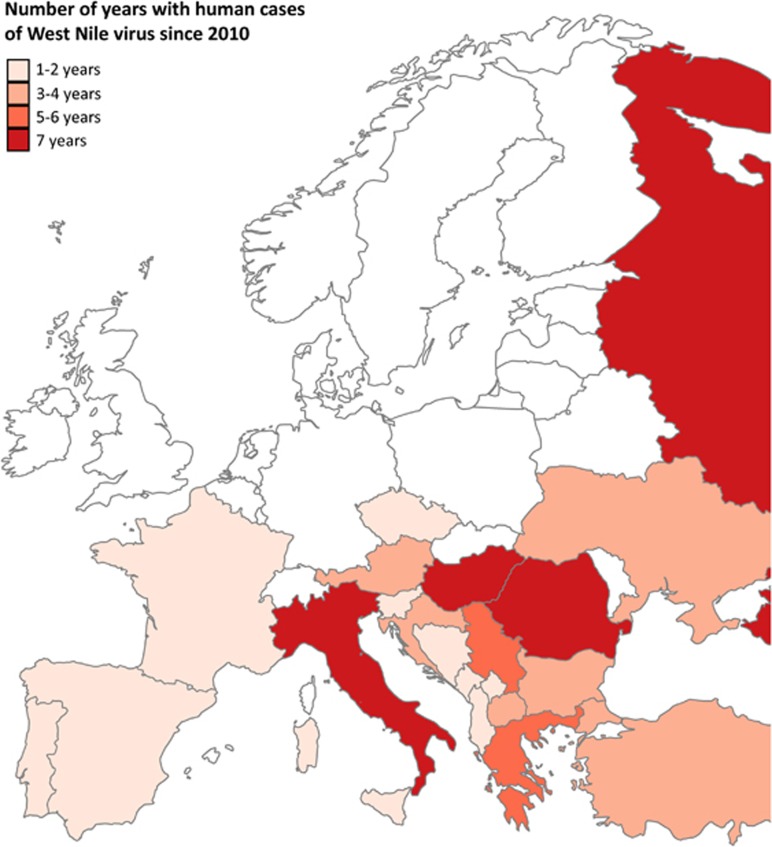
West Nile virus (WNV) is an arthropod-borne flavivirus of high medical and veterinary importance. The main vectors for WNV are mosquito species of the Culex genus that transmit WNV among birds, and occasionally to humans and horses, which are 'dead-end' hosts. Recently, several studies have been published that aimed to identify the mosquito species that serve as vectors for WNV in Europe. These studies provide insight in factors that can influence vector competence of European mosquito species for WNV. Here, we review the current knowledge on vector competence of European mosquitoes for WNV, and the molecular knowledge on physical barriers, anti-viral pathways and microbes that influence vector competence based on studies with other flaviviruses. By comparing the 12 available WNV vector competence studies with European mosquitoes we evaluate the effect of factors such as temperature, mosquito origin and mosquito biotype on vector competence. In addition, we propose a standardised methodology to allow for comparative studies across Europe. Finally, we identify knowledge gaps regarding vector competence that, once addressed, will provide important insights into WNV transmission and ultimately contribute to effective strategies to control WNV.


Figures

Figure 1
Figure 1
European countries with human cases of West Nile fever reported to the European centre for disease prevention and control (ECDC). Colour gradient indicates the number of years during which cases of West Nile virus in humans have been reported since 2010. Reported cases are marked on the country level, except for the Balearic islands, Sicily, Sardinia, and Corsica, which are individually marked. Countries with no reported cases or no data available are marked in white. Data set provided by ECDC based on the data provided by WHO and Ministries of Health from the affected countries.
Figure 2
Figure 2
Schematic overview of the mosquito barriers to arbovirus infection. Schematic longitudinal cross-section of a mosquito. Arrows indicate the passage of virions through the midgut (MG) and salivary gland (SG) barriers. The dashed circle in the midgut represents the peritrophic membrane that is formed after ingestion of blood. Right inset: (i) Infection of midgut epithelial cells via binding to a putative receptor protein. (ii) Virus replication in midgut epithelial cells. (iii) Release of virus via budding from midgut epithelial cells and direct passage through the basal lamina into the haemocoel. (iv) Direct virus passage into the haemocoel through a ‘leaky’ midgut. (v) Virus infection of trachea after budding from midgut epithelial cells. (vi) Budding of virus from the trachea into the haemocoel. Left inset: (i) Infection of the salivary gland epithelial cells after passage through the basal lamina. (ii) Virus replication in the salivary gland cells. (iii) Virus release via budding from salivary gland cells into the salivary gland lumen. (iv) Virus release from the salivary gland cells into the salivary gland lumen via apoptosis.

References

    1. Bowen RA, Nemeth NM. Experimental infections with West Nile virus. Curr Opin Infect Dis 2007; 20: 293–297. - PubMed
    1. Petersen LR, Brault AC, Nasci RS. West Nile virus: review of the literature. JAMA 2013; 310: 308–315. - PMC - PubMed
    1. Buckley A, Dawson A, Gould EA. Detection of seroconversion to West Nile virus, Usutu virus and Sindbis virus in UK sentinel chickens. Virol J 2006; 3: 71. - PMC - PubMed
    1. Buckley A, Dawson A, Moss SR et al. Serological evidence of West Nile virus, Usutu virus and Sindbis virus infection of birds in the UK. J Gen Virol 2003; 84: 2807–2817. - PubMed
    1. Linke S, Niedrig M, Kaiser A et al. Serologic evidence of West Nile virus infections in wild birds captured in Germany. Am J Trop Med Hyg 2007; 77: 358–364. - PubMed

MeSH terms

LinkOut - more resources