Review
. 2017 Nov 8;5(4):42.
doi: 10.3390/vaccines5040042.

Mechanisms of Entry and Endosomal Pathway of African Swine Fever Virus

Affiliations
Review

Mechanisms of Entry and Endosomal Pathway of African Swine Fever Virus

Elena G. Sánchez et al. Vaccines (Basel). .

Abstract

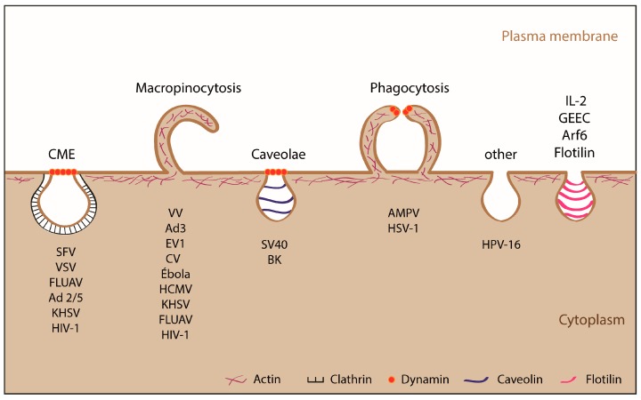
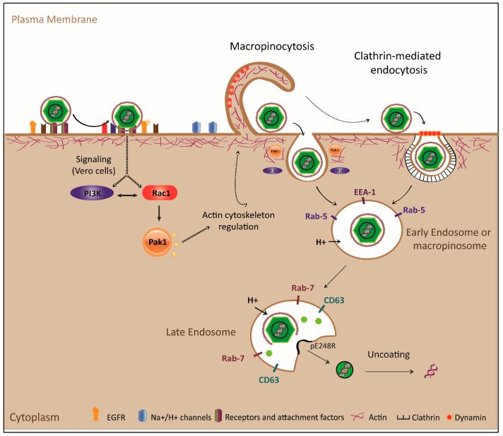
African Swine Fever Virus (ASFV) causes a serious swine disease that is endemic in Africa and Sardinia and presently spreading in Russia and neighboring countries, including Poland and recently, the Czech Republic. This uncontrolled dissemination is a world-wide threat, as no specific protection or vaccine is available. ASFV is a very complex icosahedral, enveloped virus about 200 nm in diameter, which infects several members of pigs. The virus enters host cells by receptor-mediated endocytosis that depends on energy, vacuolar pH and temperature. The specific receptor(s) and attachment factor(s) involved in viral entry are still unknown, although macropinocytosis and clathrin-dependent mechanisms have been proposed. After internalization, ASFV traffics through the endolysosomal system. The capsid and inner envelope are found in early endosomes or macropinosomes early after infection, colocalizing with EEA1 and Rab5, while at later times they co-localize with markers of late endosomes and lysosomes, such as Rab7 or Lamp 1. A direct relationship has been established between the maturity of the endosomal pathway and the progression of infection in the cell. Finally, ASFV uncoating first involves the loss of the outer capsid layers, and later fusion of the inner membrane with endosomes, releasing the nude core into the cytosol.

Keywords: ASFV; CME; PI3K; Pak-1; Rac-1; endosomes; macrophages; macropinocytosis; signaling; uncoating; viral entry.


Conflict of interest statement

The author(s) declare no competing financial interests.

Figures

Figure 1
Figure 1
Endocytic mechanisms used by animal viruses. In general viruses smaller than 150 nm internalize by clathrin-mediated endocytosis (CME) or caveolae mechanisms, whereas those larger than 150–200 nm use macropinocytosis or phagocytosis. Other mechanisms such as Arf-6, IL-2, flotilin and GPI-AP enriched early endosomal compartment (GEEG) are less commonly used by viruses to enter into cell. It is important to note that some viruses can enter the cell through more than one type of endocytosis. SFV, Semliki Forest Virus; VSV, Vesicular Stomatitis Virus; FLUAV, Influenza A Virus; Ad2/5, Adenovirus 2 and 5; KHSV, Kaposi Sarcoma-associated Herpesvirus; HIV-1, Human Immunodeficiency Virus; VV, Vaccinia Virus; Ad3, Adenovirus 3; EV1, Echovirus 1; CV, Coxsackie Virus, HCMV, Human Cytomegalovirus; SV40, Simian Vacuolating Virus 40; BK, Virus BK; AMPV, Mimivirus; HSV-1, Herpes Simplex Virus; HPV-16, Human Papillomavirus 16. Adapted from [36].
Figure 2
Figure 2
Rac1 localizes to ruffles during ASFV entry. Vero cells were transfected with EGFP-Rac-1 plasmid expression for 16 h and synchronously infected (adsorption period of 90 min at 4 °C) at an multiplicity of infection (MOI) of 10. Cells were fixed and incubated with Topro-3 and monoclonal antibody anti-p72 17LD3 to visualize DNA and viral particles, respectively; samples were processed by CLSM (x63 magnification). Images are representative of three independent experiments (images from D. Pérez-Núñez, E. G. Sanchez and Y. Revilla).
Figure 3
Figure 3
Model for ASFV entry and uncoating. Interaction of the viral particle with membrane receptors and attachment factors activates PI3K, EGFR, Rac1 and Pak-1 signaling pathways, which regulate actin dynamics, forming ruffles to internalize by macropinocytosis in Vero cells. In the case of swine macrophages, although the virus uses macropinocytosis, it does not actively induce the pathway; ASFV is also able to enter cells by CME. After viral uptake, particles are endocytosed in early endosomes or macropinosomes and transported to late endosomes where the pH-dependent uncoating process takes place. The viral outer envelope is disassembled and the inner envelope fuses with the endosomal membrane, delivering viral cores to cytosol, where viral protein pE248R plays an important role.

References

    1. Alcami A., Carrascosa A.L., Vinuela E. The entry of African swine fever virus into vero cells. Virology. 1989;171:68–75. doi: 10.1016/0042-6822(89)90511-4. - DOI - PubMed
    1. Geraldes A., Valdeira M.L. Effect of chloroquine on African swine fever virus infection. J. Gen. Virol. 1985;66:1145–1148. doi: 10.1099/0022-1317-66-5-1145. - DOI - PubMed
    1. Valdeira M.L., Geraldes A. Morphological study on the entry of African swine fever virus into cells. Biol. Cell. 1985;55:35–40. doi: 10.1111/j.1768-322X.1985.tb00407.x. - DOI - PubMed
    1. Alcami A., Carrascosa A.L., Vinuela E. Saturable binding sites mediate the entry of African swine fever virus into vero cells. Virology. 1989;168:393–398. doi: 10.1016/0042-6822(89)90281-X. - DOI - PubMed
    1. Alcami A., Carrascosa A.L., Vinuela E. Interaction of African swine fever virus with macrophages. Virus Res. 1990;17:93–104. doi: 10.1016/0168-1702(90)90071-I. - DOI - PubMed

LinkOut - more resources