Comparative Study
. 2017 Nov 8;12(11):e0187288.
doi: 10.1371/journal.pone.0187288. eCollection 2017.

Genomic comparison between Staphylococcus aureus GN strains clinically isolated from a familial infection case: IS1272 transposition through a novel inverted repeat-replacing mechanism

Affiliations
Comparative Study

Genomic comparison between Staphylococcus aureus GN strains clinically isolated from a familial infection case: IS1272 transposition through a novel inverted repeat-replacing mechanism

Tsai-Wen Wan et al. PLoS One. .

Abstract

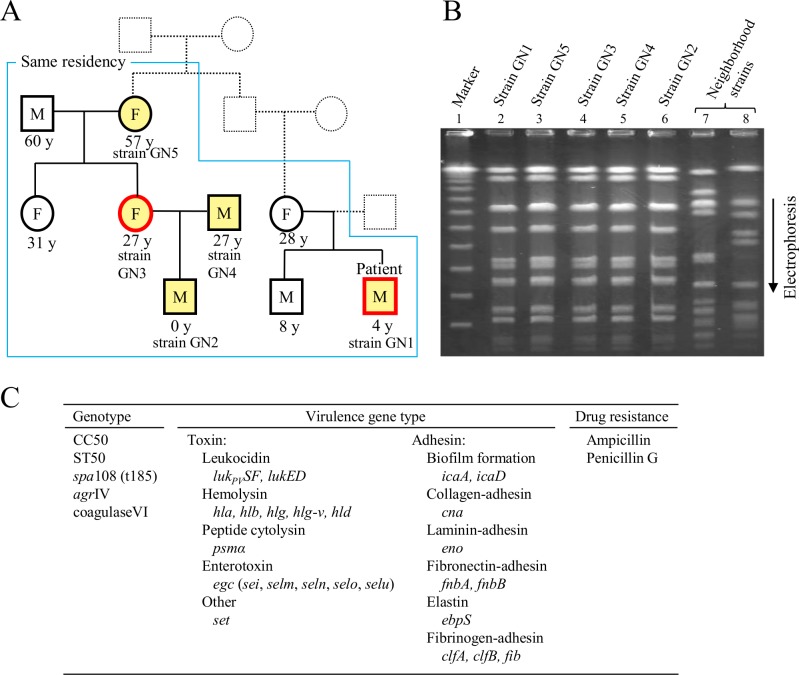
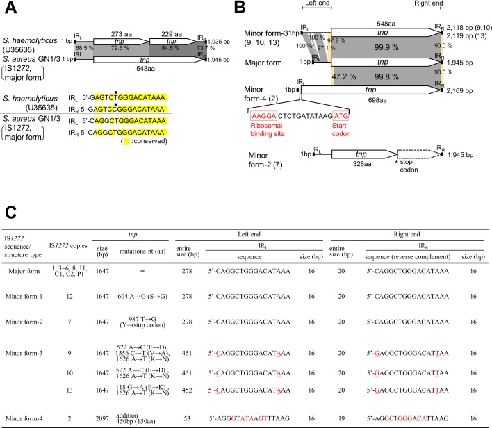
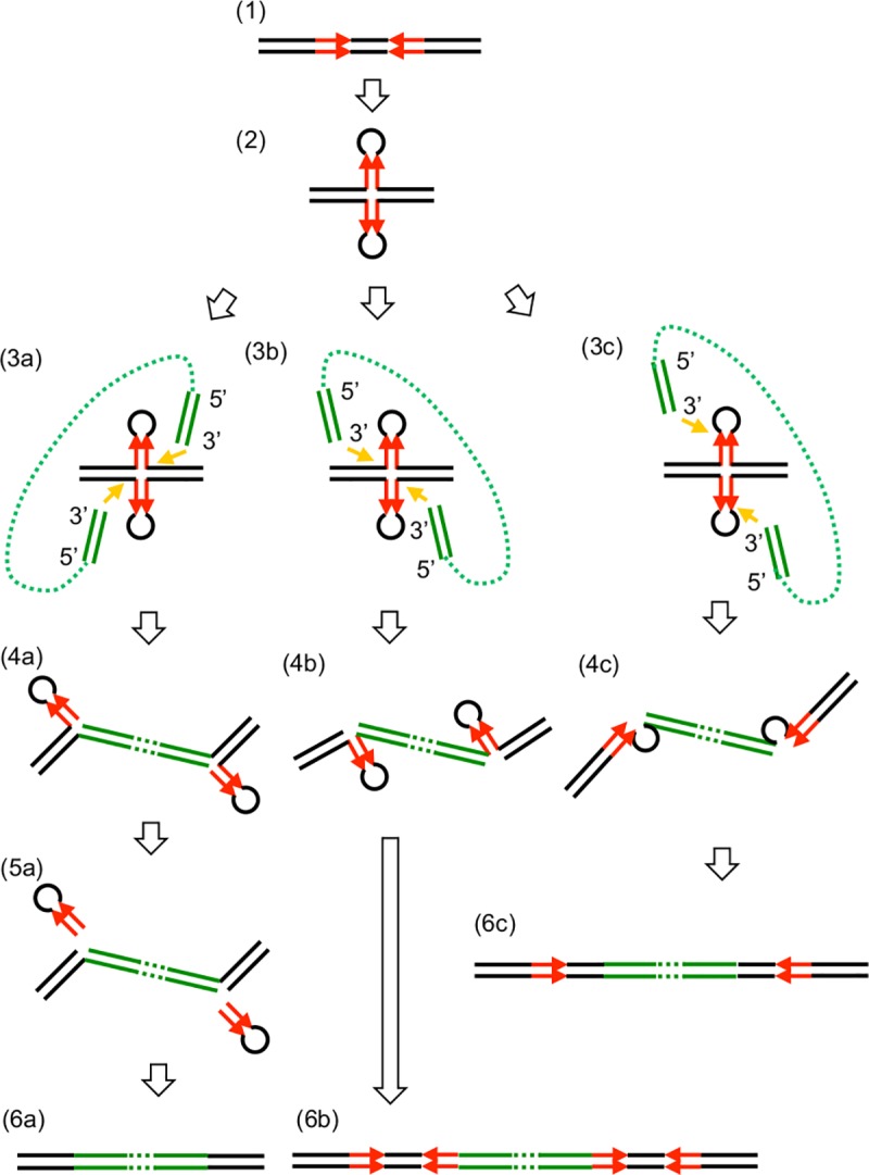
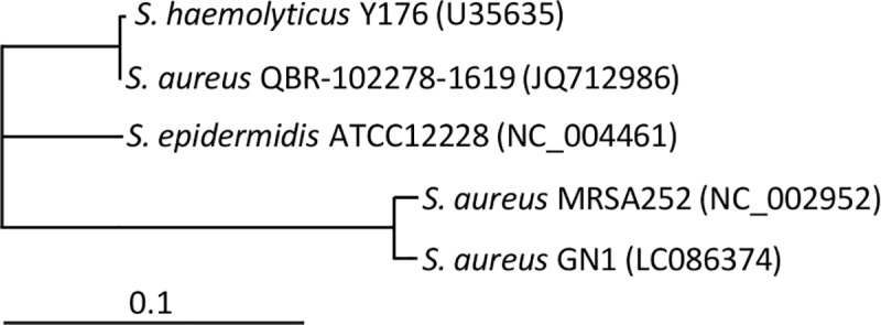
A bacterial insertion sequence (IS) is a mobile DNA sequence carrying only the transposase gene (tnp) that acts as a mutator to disrupt genes, alter gene expressions, and cause genomic rearrangements. "Canonical" ISs have historically been characterized by their terminal inverted repeats (IRs), which may form a stem-loop structure, and duplications of a short (non-IR) target sequence at both ends, called target site duplications (TSDs). The IS distributions and virulence potentials of Staphylococcus aureus genomes in familial infection cases are unclear. Here, we determined the complete circular genome sequences of familial strains from a Panton-Valentine leukocidin (PVL)-positive ST50/agr4 S. aureus (GN) infection of a 4-year old boy with skin abscesses. The genomes of the patient strain (GN1) and parent strain (GN3) were rich for "canonical" IS1272 with terminal IRs, both having 13 commonly-existing copies (ce-IS1272). Moreover, GN1 had a newly-inserted IS1272 (ni-IS1272) on the PVL-converting prophage, while GN3 had two copies of ni-IS1272 within the DNA helicase gene and near rot. The GN3 genome also had a small deletion. The targets of ni-IS1272 transposition were IR structures, in contrast with previous "canonical" ISs. There were no TSDs. Based on a database search, the targets for ce-IS1272 were IRs or "non-IRs". IS1272 included a larger structure with tandem duplications of the left (IRL) side sequence; tnp included minor cases of a long fusion form and truncated form. One ce-IS1272 was associated with the segments responsible for immune evasion and drug resistance. Regarding virulence, GN1 expressed cytolytic peptides (phenol-soluble modulin α and δ-hemolysin) and PVL more strongly than some other familial strains. These results suggest that IS1272 transposes through an IR-replacing mechanism, with an irreversible process unlike that of "canonical" transpositions, resulting in genomic variations, and that, among the familial strains, the patient strain has strong virulence potential based on community-associated virulence factors.


Conflict of interest statement

Competing Interests: The authors have declared that no competing interests exist.

Figures

Fig 1
Fig 1. Intrafamilial transmission and molecular characteristics of Panton-Valentine leukocidin (PVL)-positive Staphylococcus aureus.
(A) Nine family members (from three families) were living together with the 4-year-old boy (a patient who had skin abscesses) within the same house. Five members who were infected with PVL-positive S. aureus are marked with yellow. Four members, except the 4-year-old patient, did not develop any symptoms. Red square and circle indicate that the gonomes of PVL-positive S. aureus were sequenced. PVL-positive S. aureus srains: GN1, patient strain; GN2, infant strain; GN3, 27-year-old parent (female) strain; GN4, 27-year-old parent (male) strain; GN5, strain from the oldest member among infected members. (B) Familial strains (GN1 to GN5) were analyzed by pulsed-field gel electrophoresis, suggesting a clonal infection from one PVL-positive S. aureus source. Neighborhood strains were from healthy persons from neighboring families unrelated to the patient's families. (C) The genotypes, vurulence genes analyzed by PCR, and drug resistance of familial strains GN1 to GN5 are summarized, indicating the common features among strains GN1 to GN5.
Fig 2
Fig 2. Circular genome maps of familial strains GN1 and GN3.
Genomes: A, GN1; B, GN3. Genome information includes S. aureus-typing targets, phages, mobile genetic elements, including IS1272, deletion, virulence, and drug resistance. Genes (products) described on the genome map are: spa, protein A (IgG-binding protein); coa, coagulase; psma, phenol-soluble modulin α(PSMα, cytolytic peptide); gltA, glutamate synthase; fib, fibrinogen adhesin; hla, α-hemolysin (Hla); ebhA, extracellular matrix-binding protein/very large surface-anchored protein/giant protein (Ebh); rot, repressor of toxins; lukE-lukD, bi-component leukocidin; egc, enterotoxin gene cluster carrying sei, selm, seln, selo, and selu; map, map protein; hlb, β-hemolysin (Hlb); hld, δ-hemolysin (Hld, cytolytic peptide); agr, accessory gene regulator; gltT, proton/sodium-glutamate symport protein, sbi, second binding protein of immunoglobulin, hlg, γ-hemolysin (Hlg); fnb, fibronectin-binding protein; ica, intercellular adhesion protein A (biofilm formation); cna, collagen adhesin. The DNA helicase gene and eight other helicase genes, dnaB, recQ1, comFA, recG, recQ, ruvB, rubA, and dnaB2, were also mapped on the genomes; they are shown in purple. GN1 and GN3 had PVL-converting φSa2 (φPVL-Sa2GN1 and φPVL-Sa2GN3, respectively), carrying the PVL genes (lukPVSF). Unique genetic structures were carried by φSa3 remnant, the immune evasion cluster (IEC), composed of three immune evasion genes, sak (staphylokinase, SAK), chp (chemotaxis inhibitory protein of S. aureus, CHIPS), and scn (staphylococcal complement inhibitor, SCIN), being present on the GN genome as IEC/PR. Penicillin resistance was encoded by a chromosomal bla-tnp structure, carrying an array of blaI-blaR1-blaZ and tnpC-tnpB-tnpA. The GN1 and GN3 genomes carried 14 and 15 copies of IS1272, respectively. Of those, 13 copies were commonly-existing IS1272 (ce-IS1272), and named 1 to 13; moreover, GN1 had one copy of newly-inserted IS1272 (ni-IS1272), which was named P1, while GN2 had two copies of ni-IS1272, which were named C1 and C2, as shown in A and B, respectively. ce-IS1272 copy 6 and ni-IS1272 copy C1 formed a larger structure (designated as IS1272L). ce-IS1272 copy 2, labeled in green, had the transposase gene (tnp) encoding for a long fusion form of transposase; ce-IS1272 copy 7, marked with an asterisk, had tnp encoding for a truncated form of transposase. The direction of the IS1272 insertion is shown by + or -. The targets of IS1272 transposition are indicated by color: red, IRs; blue, “non-IRs”; yellow and green, unknown (target sequences are too big in size). The 3-kb region, located close to IS1272 copy 3, in GN1 (A) was deleted in GN3 (B).
Fig 3
Fig 3
Comparison of IS1272 from S. haemolyticus and S. aureus GN (A), and comparison between IS1272 copies on the GN genomes (B and C). (A) The IS1272 of S. haemolyticus has two transposase (Tnp) genes (ORFs, tnp) and heterogenous IRs (sequence identity, 15 of 16 bp); divergent nucleotides are shown by a dot [33]. In contrast, a major form of IS1272 in GN1 and GN3 had only one tnp and homogeneous IRs (sequence identity, 16 of 16 bp). (B) The structures of IS1272, major form and minor forms (2, 3, and 4), were compared. Homologous regions are shaded in each comparison. IS1272/minor form-2 (copy 7) had a premature stop codon in tnp, thus its product was predicted to be truncated Tnp. IS1272/minor form-4 (copy 2) had tnp of a larger size, which started at an ATG codon located upstream of tnp/major form; the ribosome binding sequence (AAGGA), which can potentially pair with the complementary sequence at the 3'-end of 16S rRNA [47], is shown in red. (C) The genetic statuses of 16 IS1272 copies on the GN1/GN3 genomes are summarized. IS1272/major form had 1,647-bp (548-aa) tnp and 16-bp homogeneous IRs (sequence identity, 16 of 16 bp). IS1272/minor form-1 had a nonsynonymous substitution in tnp. IS1272/minor form-2 had a premature stop codon in tnp. IS1272/minor form-3 had 16-bp heterogeneous IRs (sequence identity, 14 of 16 bp) and tnp which was divergent in comparison with tnp/major form. IS1272/minor form-4 had 16-bp more-divergent IRs (sequence identity, 10 of 16 bp) and tnp with a 450-bp (150-aa) longer N-terminal side. nt, nucleotide.
Fig 4
Fig 4. Structure of IS1272 with tandem duplications of its left side sequence.
ce-IS1272 copy 6 in GN1 and GN3 and ni-IS1272 copy C1 in GN3 formed a larger IS1272 structure (designated as IS1272L) with the same sequence. IS1272L had tandem duplications of the 25-bp left side sequence including IRL; the size of IS1272L with IRL2 and IRR is 1,970 bp.
Fig 5
Fig 5. Evidence of inverted repeats (IR)-replacing transposition for IS1272.
Comparison of GN1 and GN3 genome sequences at a position of newly-inserted IS1272 (ni-IS1272) made it possible to definitely assign a set of target and IS1272 insertion. Color: red, target inverted repeats (IRs); yellow, terminal IRs of IS1272; blue, the same DNA sequence region between GN1 and GN3. In A, IS1272 transposition occurred on the PVL-prophage, targeting a 24-bp sequence with 9-bp IRs and yielding IS1272-P1 (on GN1). In B, IS1272 transposition occurred in a region downstream of rot (and also downstream of the rRNA methyltransferase gene), targeting a 30-bp sequence with 12-bp IRs and yielding IS1272-C1 (on GN3). In C, IS1272 transposition occurred within the DNA helicase gene, targeting a 22-bp sequence with 9-bp IRs and yielding the larger IS1272 structure (IS1272L) of IS1272-C1 (on GN3); this IS1272L may have occurred by the tandem duplication of the 25-bp left side sequence upon transposition or by transposition of IS1272L. In all the three cases, the target IRs are replaced with IS1272.
Fig 6
Fig 6. Analysis of previous transposition modes for current IS1272 copies.
For 13 copies of commonly-existing IS1272 (ce-IS1272) on the GN1 and GN3 genomes, possible targets of transposition were searched in the database. Color: red, target inverted repeats (IRs); yellow, IS1272 with terminal IRs; blue, the same DNA sequence region between the GN1/GN3 genome and the genome searched in the database. In A, the possible targets were IRs for seven copies of ce-IS1272 (copies 1, 3, 4, 7–10). In B, the possible targets were “non-IRs” for three copies of ce-IS1272 (copies 6, 11, 12). For the remaining three copies of ce-IS1272 (copies 2, 5, 13), the possible targets were unknown (target sequences not specific or too big in size).
Fig 7
Fig 7. Target inverted repeat (IR) sequence analysis for IS1272 on the GN genomes.
For newly-inserted IS1272 (ni-IS1272), target site sequences were analyzed by comparing the GN1 and GN3 genome sequences at each ni-IS1272 insertion site. For commonly-existing IS1272 (ce-IS1272), target site sequences were searched from the database. IRs in the target site sequences are underlined. The target site sequences exhibited no sequence homology to each other, suggesting the role of a stem-loop structure as a target of IS1272 transposition.
Fig 8
Fig 8. Target inverted repeat (IR) sequence analysis using target/IS1272 sets obtained in database searches.
The IR sequences from 19 target/IS1272 sets were searched using the database and are summarized in figures. Those for IS1272 copy P1 are from the GN1/GN3 genomes. Terminal IRs in the target sequences are underlined. IS1272 IR sequences with red nucleotides represent heterogeneous IRs; red nucleotides are divergent in the left and right IRs. Model A shows the results when the size of IRs is 16 bp; model B shows the results when the size of IRs is 16 bp or more.
Fig 9
Fig 9. Variation in size of the IS1272 transposase (Tnp) gene (tnp).
(A) The sizes of the IS1272 tnp genes in GN1/GN3, other reported S. aureus (including MRSA), and S. hemolyticus are summarized in figures; other S. aureus included strains 6850, MRSA252, TCH60, JKD6159, SA268, T-ORC_001, SA957, and DAR4145 (they were from the database) and S. haemolyticus was from [33]. The majority of tnp genes encoded for a 548-aa product. However, for example, GN1/3 copy 7 tnp had a premature stop codon, resulting in a truncated product (328 aa); and S. haemolyticus IS1272 tnp had one base deletion, resulting in a frame shift mutation and two smaller ORFs (229 aa and 273 aa), due to one more deletion [31]. Moreover, GN1/3 copy 2 tnp encoded for a larger product (698 aa). (B) This figure shows that the tnp gene of GN1/3 IS1272 copy 2 encodes for a fusion protein, constructed by isochorismatase (putative), shown in red, and GN1 IS1272 copy P1 transposase, shown in blue. There was a link peptide region (PI), shown in black, between the isochorismatase (putative) domain and IS1272 P1 transposase domain; the same link peptide region was also present in both isochorismatase (putative) and IS1272 P1 transposase; the nucleotide sequence corresponding to the link peptide region was 5’-CCAATA.
Fig 10
Fig 10. Unique genetic structures on the GN1 and GN3 genomes.
(A) The GN3 genome, but not GN1 genome, had IS1272 insertion within the DNA helicase gene, yielding newly-inserted IS1272 (ni-IS1272) copy C1. The copy C1 formed a larger IS1272 structure (IS1272L) with a tandem duplication of the 25-bp left side sequence including IRL. Due to the IS1272 C1 insertion, particularly the internal stop codon TAA which was present in IRL2 of IS1272L, the DNA helicase gene product was estimated to be truncated (973-aa) DNA helicase. (B) The GN3 genome had a small (3-kb) deletion close to IS1272 (copy 3); the 3-kb region was present on the GN1 genome and also on the genome of related ST50 MSSA strain 6850. (C) The immune evasion cluster (IEC), carrying sak for staphylokinase (SAK), chp for chemotaxis inhibitory protein of S. aureus (CHIPS), and scn for staphylococcal complement inhibitor (SCIN), was carried by the φSa3 remnant. This structure, IEC/PR, had the att of φSa3; 5 out of 13 nucleotides were divergent (divergent nucleotides are underlined). Its location was not hlb (insertion site of φSa3). GN1 and GN3 lacked φSa3, and hlb was intact. The IEC/PR structure carried one IS1272 copy (copy 7). (D) The blaZ,R1,I-tnpA,B,C structure encodes for penicillin resistance. This structure is flanked by short (6-bp) direct repeats. This structure also carried one IS1272 copy (copy 11).
Fig 11
Fig 11. Panton-Valentine leukocidin (PVL) production levels of familial strains GN1 to GN5.
Bacteria were grown in a liquid medium for 18 h with or without 5% fetal bovine serum (FBS), serial doubling dilutions of the culture supernatants were made, and the amounts of PVL in the supernatants were serologically measured. Bars (color): black, PVL production in the absence of FBS; red, PVL production in the presence of FBS. The PVL production levels of GN1 and GN2 were two-fold higher than those of GN3, GN4, and GN5. Addition of serum to the bacterial culture medium resulted in two fold-higher PVL production levels in any case. USA300-0114 was used as a PVL-positive control strain.
Fig 12
Fig 12. mRNA expression levels of cytolytic peptide genes (psmα and hld) and Panton-Valentine leukocidin (PVL) genes (lukPVSF) in familial strains GN1 to GN5.
Bacteria were grown on sheep blood agar for 8 h, and the mRNA expression levels were examined by an RT-PCR assay. PVL-positive CA-MRSA USA300-0114 was used as a control strain which shows high expression levels for psmα and hld, and PVL-negative HA-MRSA Mu50 was used as a control strain which shows low expression levels for psmα and hld. In (A), the products in an RT-PCR assay were visualized on 2% agarose gels after electrophoresis. In (B), the expression data of each strain were normalized to those of USA300-0114. For psmα: P1 vs. P2, P<0.05; P3 vs. P2, P<0.05; P4 vs. P2, P<0.05; P3 vs. P4 (group of GN2, GN3, GN4, and GN5), P<0.05. For hld, H1 vs. H2, P<0.05; H3 vs. H2, P<0.05; H4 vs. H2, P<0.05; H3 vs. H4 (group of GN2, GN3, GN4, and GN5), P<0.05. For PVL genes: L1 (group of GN1 and GN2) vs. L2 (group of GN3, GN4, and GN5), P<0.05. The LVL mRNA expression level of GN5 was unexpectedly low, compared with that of other familial strains (P<0.05). PVL-positive CA-MRSA strain RS08 and CA-MSSA strain KT1 were also used as strong psmα/hld expression control strains, the data being comparable to that of CA-MRSA USA300-0114 (for psmα, 1.00, 1.09, and 0.92 for USA300-0114, RS08, and KT1, respectively; and for hld, 1.00, 0.85, and 0.79 for USA300-0114, RS08, and KT1, respectively); and HA-MRSA strain N315 was also used as a low psmα/hld expression control strain, the data being comparable to that of HA-MRSA Mu50 (for psmα, 0.45 and 0.38 for Mu50 and N315, respectively; and for hld, 0.41 and 0.30 for Mu50 and N315, respectively).
Fig 13
Fig 13. Replacement by structure-dependent transposition (RST) or stem-loop replacement: A possible model for the transposition mechanism of IS1272.
(1) Palindromic sequences (red) are present in donor and recipient sequences. (2) Palindromic sequences form a cruciform. (3a-4a) The 3’ ends of IS1272 (stem-loop) attack and are joined to the target DNA at the 3’ feet of two stem-loops. (5a) The stem-loops on recipient sequences are removed. (6a) IS is inserted, replacing a stem-loop sequence. (3b-4b) The 3’ ends of IS attack and are joined to the target DNA at the 5’ feet of the two stem-loop. (6b) The sequence forming the stem-loop is duplicated at both ends of IS. (3c-4c) The 3’ ends of IS attack and are joined to target DNA at the 5’ of two loops. (6c) The sequence corresponding to the two loops is duplicated at both ends of IS. Red arrows, palindromic sequences; green lines, IS DNA.
Fig 14
Fig 14. Cluster analysis of IS1272 from S. aureus, S. epidermidis, and S. haemolyticus.
GenBank accession numbers are shown in parentheses.

References

    1. Tacconelli E, Tumbarello M, Cauda R. Staphylococcus aureus infections. N Engl J Med. 1998; 339,2026–2027. - PubMed
    1. Tenover FC, Gorwitz RJ. The epidemiology of Staphylococcus infections In: Fischetti VA, Novick RP, Ferretti JJ, Portnoy DA, Rood JI, editors. Gram-positive pathogens, 2nd Ed. Washington, DC: ASM Press; 2006. pp. 526–534.
    1. Powers ME, Bubeck Wardenburg J. Igniting the fire: Staphylococcus aureus virulence factors in the pathogenesis of sepsis. PLoS Pathog. 2014; 10:e1003871 doi: 10.1371/journal.ppat.1003871 - DOI - PMC - PubMed
    1. Kobayashi SD, Malachowa N, DeLeo FR. Pathogenesis of Staphylococcus aureus abscesses. Am J Pathol. 2015; 185:1518–1527. doi: 10.1016/j.ajpath.2014.11.030 - DOI - PMC - PubMed
    1. Tong SY, Davis JS, Eichenberger E, Holland TL, Fowler VG Jr. Staphylococcus aureus infections: epidemiology, pathophysiology, clinical manifestations, and management. Clin Microbiol Rev. 2015; 28,603–661. doi: 10.1128/CMR.00134-14 - DOI - PMC - PubMed

Publication types

MeSH terms