. 2017 Dec 1;24(6):585-596.
doi: 10.1093/dnares/dsx027.

Sequencing and phasing cancer mutations in lung cancers using a long-read portable sequencer

Affiliations

Sequencing and phasing cancer mutations in lung cancers using a long-read portable sequencer

Ayako Suzuki et al. DNA Res. .

Abstract

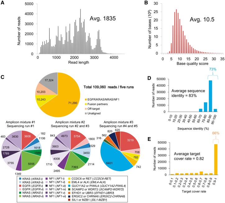
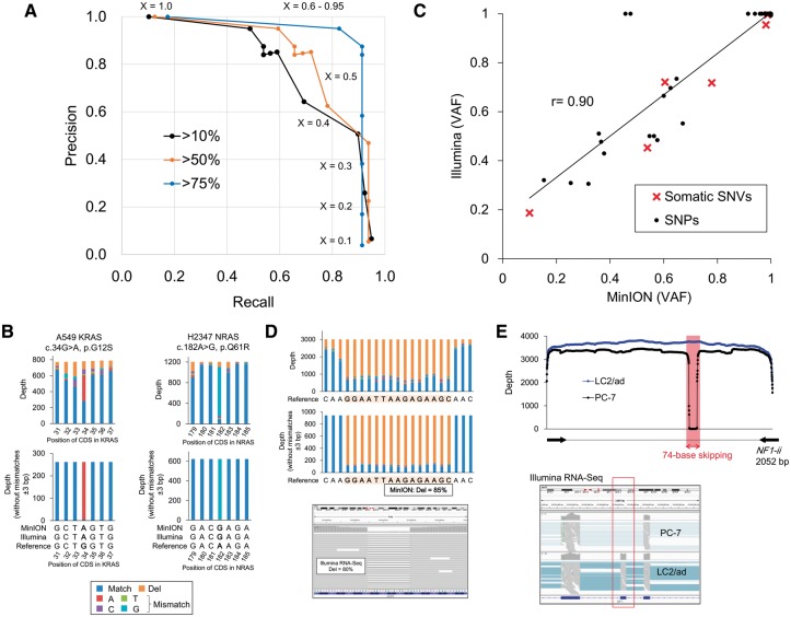
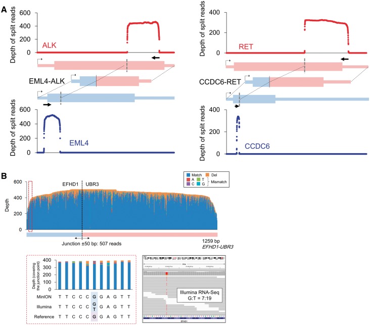
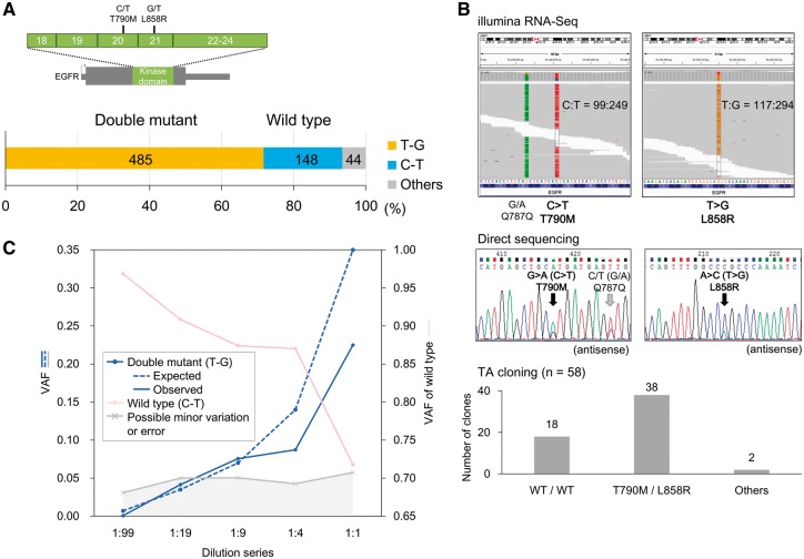
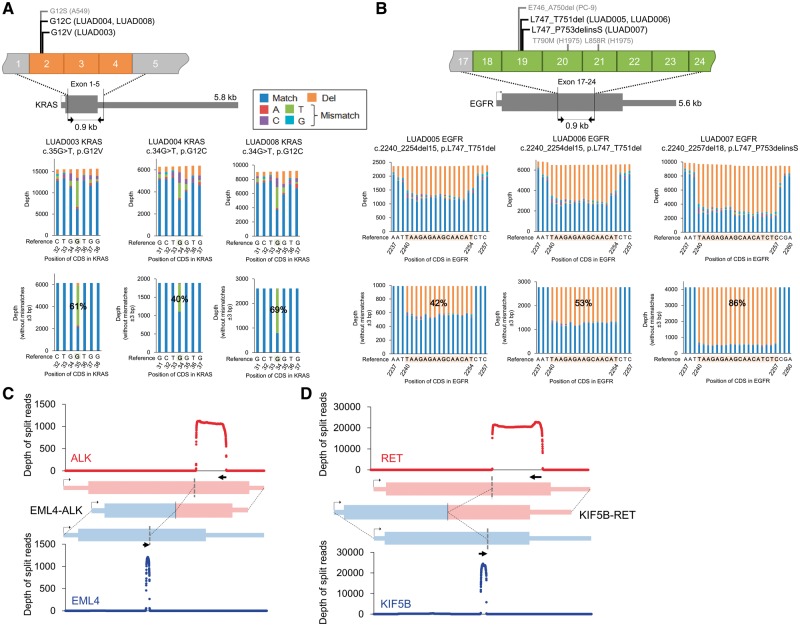
Here, we employed cDNA amplicon sequencing using a long-read portable sequencer, MinION, to characterize various types of mutations in cancer-related genes, namely, EGFR, KRAS, NRAS and NF1. For homozygous SNVs, the precision and recall rates were 87.5% and 91.3%, respectively. For previously reported hotspot mutations, the precision and recall rates reached 100%. The precise junctions of EML4-ALK, CCDC6-RET and five other gene fusions were also detected. Taking advantages of long-read sequencing, we conducted phasing of EGFR mutations and elucidated the mutational allelic backgrounds of anti-tumor drug-sensitive and resistant mutations, which could provide useful information for selecting therapeutic approaches. In the H1975 cells, 72% of the reads harbored both L858R and T790M mutations, and 22% of the reads harbored neither mutation. To ensure that the clinical requirements can be met in potentially low cancer cell populations, we further conducted a serial dilution analysis of the template for EGFR mutations. Several percent of the mutant alleles could be detected depending on the yield and quality of the sequencing data. Finally, we characterized the mutation genotypes in eight clinical samples. This method could be a convenient long-read sequencing-based analytical approach and thus may change the current approaches used for cancer genome sequencing.

Keywords: MinION; cancer mutations; lung cancer cell lines; phasing.


Figures

Figure 1
Figure 1
Summary of the amplicon sequencing and alignment statistics. (A, B) Distribution of the read lengths (A) and QVs (B) in all five sequencing runs. The average number is shown in the inset. (C) The number of MinION reads aligned to each of the PCR target regions. For the alignment, we used LAST with tuned parameters as described in the Methods section. (D, E) Distributions of the sequence identity (D) and target cover rate (E) in each read. The average number is shown in the inset.
Figure 2
Figure 2
Detection of SNPs and mutations in the MinION reads. (A) Precision and recall rates of SNV detection using MinION. Blue, orange and black lines represent three datasets of SNVs corresponding to different variant allele frequencies (VAF) of Illumina standards, which are more than 75% (targets are only homozygous variants), 50% (targets include heterozygous variants) and 10% (targets include minor population variant). ‘X’ represents one of the parameters for the SNV detection, which is the threshold of the VAFs of the MinION reads. Additional details regarding the procedure are described in Supplementary Fig. 2. (B) The depths and base patterns of KRAS G12S in the A549 cells (left) and NRAS Q61R in the H2347 cells (right). The pre-cleaned data are shown in the upper panel. The cleaned data in which the MinION reads without mismatches ±3 bp of the SNVs were used are shown in the lower panel. The color key for the base patterns is represented in the margin. (C) VAFs for the Illumina RNA-Seq and MinION sequencing at the 41 SNVs. SNPs and somatic SNVs are shown as black circles and red crosses, respectively. (D) The depths and base patterns of the 15-base EGFR deletion in the PC-9 cells in the MinION reads. The pre-cleaned and cleaned data are shown in the upper and middle panel. IGV visualization of the Illumina RNA-Seq data is represented in the lower panel. The color key is the same as that shown in B. (E) Exon skipping in exon 19 of NF1 in the PC-7 cells. In the upper panel, the sequence depths of the MinION reads aligned to the NF1-ii region with split alignment using LAST are shown in the PC-7 (black) and LC2/ad (blue, wild-type) cells. The exon skipping in the Illumina RNA-Seq data is also shown in the lower panel.
Figure 3
Figure 3
Sequencing the fusion transcripts using the MinION reads.(A) Sequence depths of EML4-ALK and CCDC6-RET. The reads were split to both fusion partners with split alignment. (B) Allelic relevance between the SNP and the junction point of the EFHD1-UBR3 fusion transcript in the PC-9 cells. In the upper panel, the depths and base patterns of the MinION reads are shown in the EFHD1-UBR3 target region. The junction point is shown as a broken black line. One of the heterozygous SNPs in EFHD1, which is encircled with a red broken line and is referred to as G, co-occurred with the fusion junction in the MinION reads (left in the lower panel). This SNP was verified as heterozygous using Illumina RNA-Seq (right in the lower panel).
Figure 4
Figure 4
Phasing cancer mutations. (A) Phasing of the EGFR mutations T790M and L858R in H1975 cells. The number of MinION reads called for each of the SNV patterns. The MinION reads without mismatches ±3 bp of the positions of both SNVs. (B) T790M and L858R mutations using Illumina RNA-Seq and Sanger sequencing. Both SNVs were called as heterozygous mutations by Illumina RNA-Seq and direct Sanger sequencing in the upper and middle panels, respectively. The pattern and variant tag frequencies of both SNVs in each DNA molecule were validated with TA cloning, followed by Sanger sequencing (lower panel). (C) Variant allele frequencies (VAFs) in the five-point EGFR-mutant dilution series. The expected and observed VAFs of the phased double mutant (T790M/L858R) are shown in blue solid and dashed lines, respectively. The VAFs in the wild-type and other patterns are also represented in pink and gray, respectively.
Figure 5
Figure 5
Detection of driver alterations in clinical samples. (A, B) Detection of driver alterations in clinical samples using MinION sequencing. KRAS (A) and EGFR (B) mutations are shown for the mutation-positive patients. The PCR target regions are shown in the upper panel. The pre-cleaned and cleaned (without mismatches ±3 bp of the mutation) depths are shown in the middle and lower panel. (C, D) Sequence depths of split alignment for EML4-ALK (C) and KIF5B-RET (D).

References

    1. The Cancer Genome Atlas Research Network, Weinstein J.N., Collisson E.A., et al. 2013, The Cancer Genome Atlas Pan-Cancer analysis project. Nat. Genet., 45, 1113–20. - PMC - PubMed
    1. International Cancer Genome Consortium 2010, International network of cancer genome projects. Nature, 464, 993–8. - PMC - PubMed
    1. Sharma S.V., Bell D.W., Settleman J., Haber D.A.. 2007, Epidermal growth factor receptor mutations in lung cancer. Nat. Rev. Cancer, 7, 169–81. - PubMed
    1. Soda M., Choi Y.L., Enomoto M., et al. 2007, Identification of the transforming EML4-ALK fusion gene in non-small-cell lung cancer. Nature, 448, 561–6. - PubMed
    1. Kwak E.L., Bang Y.J., Camidge D.R., et al. 2010, Anaplastic lymphoma kinase inhibition in non-small-cell lung cancer. N. Engl J. Med., 363, 1693-1703. - PMC - PubMed

MeSH terms