

Lower cardiac index levels relate to lower cerebral blood flow in older adults
- PMID: 29117962
- PMCID: PMC5719926
- DOI: 10.1212/WNL.0000000000004707
Lower cardiac index levels relate to lower cerebral blood flow in older adults
Abstract
Objective: To assess cross-sectionally whether lower cardiac index relates to lower resting cerebral blood flow (CBF) and cerebrovascular reactivity (CVR) among older adults.
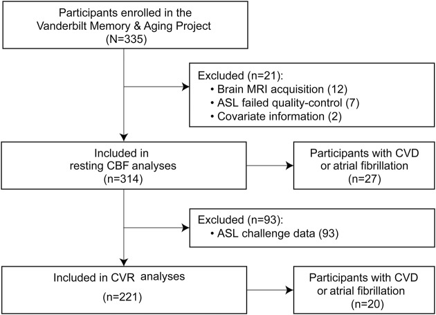
Methods: Vanderbilt Memory & Aging Project participants free of stroke, dementia, and heart failure were studied (n = 314, age 73 ± 7 years, 59% male, 39% with mild cognitive impairment). Cardiac index (liters per minute per meter squared) was quantified from echocardiography. Resting CBF (milliliters per 100 grams per minute) and hypercapnia-induced CVR were quantified from pseudo-continuous arterial spin-labeling MRI. Linear regressions with ordinary least-square estimates related cardiac index to regional CBF, with adjustment for age, education, race/ethnicity, Framingham Stroke Risk Profile score (systolic blood pressure, antihypertensive medication use, diabetes mellitus, current cigarette smoking, left ventricular hypertrophy, prevalent cardiovascular disease [CVD], atrial fibrillation), APOE ε4 status, cognitive diagnosis, and regional tissue volume.
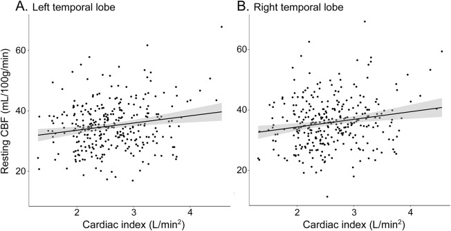
Results: Lower cardiac index corresponded to lower resting CBF in the left (β = 2.4, p = 0.001) and right (β = 2.5, p = 0.001) temporal lobes. Results were similar when participants with prevalent CVD and atrial fibrillation were excluded (left temporal lobe β = 2.3, p = 0.003; right temporal lobe β = 2.5, p = 0.003). Cardiac index was unrelated to CBF in other regions assessed (p > 0.25) and CVR in all regions (p > 0.05). In secondary cardiac index × cognitive diagnosis interaction models, cardiac index and CBF associations were present only in cognitively normal participants and affected a majority of regions assessed with effects strongest in the left (p < 0.0001) and right (p < 0.0001) temporal lobes.
Conclusions: Among older adults without stroke, dementia, or heart failure, systemic blood flow correlates with cerebral CBF in the temporal lobe, independently of prevalent CVD, but not CVR.
© 2017 American Academy of Neurology.
Figures
References
-
- Williams LR, Leggett RW. Reference values for resting blood flow to organs of man. Clin Phys Physiol Meas 1989;10:187–217. - PubMed
MeSH terms
Substances
Grants and funding
- UL1 TR000445/TR/NCATS NIH HHS/United States
- R01 NS097763/NS/NINDS NIH HHS/United States
- K23 HL128928/HL/NHLBI NIH HHS/United States
- K23 AG048347/AG/NIA NIH HHS/United States
- R01 NS078828/NS/NINDS NIH HHS/United States
- T32 EB014841/EB/NIBIB NIH HHS/United States
- K01 AG049164/AG/NIA NIH HHS/United States
- K24 AG046373/AG/NIA NIH HHS/United States
- R01 NS100980/NS/NINDS NIH HHS/United States
- R01 AG034962/AG/NIA NIH HHS/United States
- T32 HL007411/HL/NHLBI NIH HHS/United States
- K12 HL109019/HL/NHLBI NIH HHS/United States
- K23 AG045966/AG/NIA NIH HHS/United States
- S10 OD023680/OD/NIH HHS/United States
- K12 HD043483/HD/NICHD NIH HHS/United States
- L30 AG048601/AG/NIA NIH HHS/United States
LinkOut - more resources
Full Text Sources
Other Literature Sources
Miscellaneous