Review
. 2018 Feb 1;314(2):C211-C227.
doi: 10.1152/ajpcell.00022.2017. Epub 2017 Nov 8.

Carbon monoxide in lung cell physiology and disease

Affiliations
Review

Carbon monoxide in lung cell physiology and disease

Stefan W Ryter et al. Am J Physiol Cell Physiol. .

Abstract

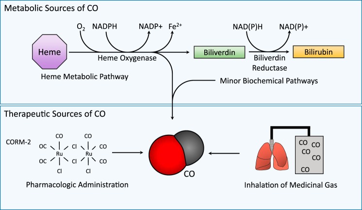
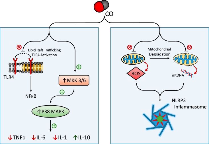
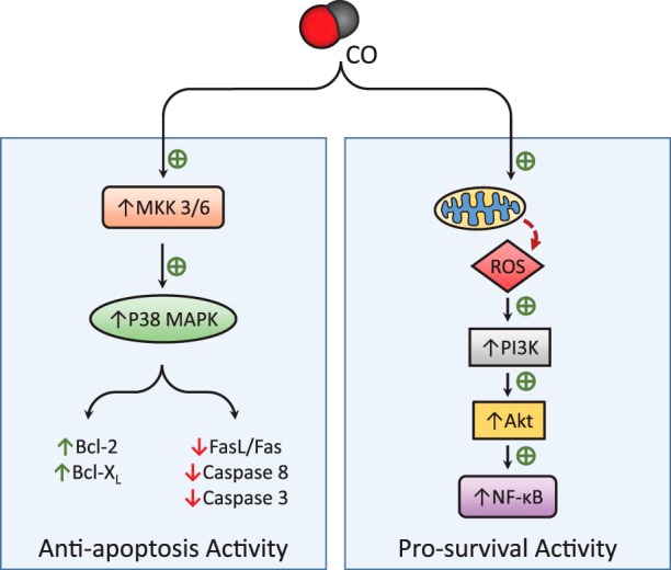
Carbon monoxide (CO) is an endogenously produced gas that has gained recognition as a biological signal transduction effector with properties similar, but not identical, to that of nitric oxide (NO). CO, which binds primarily to heme iron, may activate the hemoprotein guanylate cyclase, although with lower potency than NO. Furthermore, CO can modulate the activities of several cellular signaling molecules such as p38 MAPK, ERK1/2, JNK, Akt, NF-κB, and others. Emerging studies suggest that mitochondria, the energy-generating organelle of cells, represent a key target of CO action in eukaryotes. Dose-dependent modulation of mitochondrial function by CO can result in alteration of mitochondrial membrane potential, mitochondrial reactive oxygen species production, release of proapoptotic and proinflammatory mediators, as well as the inhibition of respiration at high concentration. CO, through modulation of signaling pathways, can impact key biological processes including autophagy, mitochondrial biogenesis, programmed cell death (apoptosis), cellular proliferation, inflammation, and innate immune responses. Inhaled CO is widely known as an inhalation hazard due to its rapid complexation with hemoglobin, resulting in impaired oxygen delivery to tissues and hypoxemia. Despite systemic and cellular toxicity at high concentrations, CO has demonstrated cyto- and tissue-protective effects at low concentration in animal models of organ injury and disease. These include models of acute lung injury (e.g., hyperoxia, hypoxia, ischemia-reperfusion, mechanical ventilation, bleomycin) and sepsis. The success of CO as a candidate therapeutic in preclinical models suggests potential clinical application in inflammatory and proliferative disorders, which is currently under evaluation in clinical trials.

Keywords: carbon monoxide; cell death; cell signaling; inflammation; lung disease; mitochondria; reactive oxygen species; sepsis.


Figures

Fig. 1.
Fig. 1.
Interaction of carbon monoxide (CO) with biological systems. CO is endogenously produced in the body, primarily as the product of the erythrocyte-dependent turnover of hemoglobin-derived heme, and the cellular turnover of hemoproteins. Heme, a catalytic cofactor for hemoproteins, is degraded by the heme oxygenase (HO, EC: 1:14:99:3) enzymes. HO catalyzes the rate-limiting step in oxidative heme catabolism, to yield biliverdin-IXα, CO, and ferrous iron (Fe II), and requires 3 mol molecular oxygen (O2) and reducing equivalents from NADPH. Biliverdin-IXα produced in the HO reaction is reduced to bilirubin-IXα by NAD(P)H biliverdin reductase (BVR). Non-heme sources may make a minor contribution to exogenous CO production. In the blood, CO binds hemoglobin to form carboxyhemoglobin (CO-Hb). Therapeutic CO may be applied at low concentration through inhalation or mechanical ventilation, or by administering CO-releasing molecules (CORMs).
Fig. 2.
Fig. 2.
Effect of CO on cellular inflammation pathways. CO has been implicated in the downregulation of inflammatory pathways. CO can inhibit the production of the proinflammatory cytokines (IL-1β, IL-6, TNF-α) and upregulate anti-inflammatory cytokine IL-10, through a mechanism dependent on activation of the MKK3/p38 MAPK pathway. Additionally, CO has been shown to modulate the trafficking and activation of Toll-like receptor-4 (TLR4) at the plasma membrane. CO has been proposed as an inhibitor of NOD-, leucine rich region- and pyrin domain-containing-3 (NLRP3) inflammasome activation, through the stabilization of mitochondria, leading to modulation of mitochondrial reactive oxygen species (mtROS) and inhibition of mtDNA release, key triggers for inflammasome activation under proinflammatory conditions.
Fig. 3.
Fig. 3.
Effect of CO on cell death pathways. CO can modulate apoptotic signaling and prosurvival pathways leading to cytoprotection. CO-dependent antiapoptotic effects have been related to the upregulation of the MKK3/p38 MAPK pathway, resulting in modulation of the expression and activation of apoptotic effector molecules. CO may target mitochondria, leading to modulation of mtROS flux and downstream adaptive signaling. Furthermore, the stimulation of phosphoinositide 3-kinase (PI3K)/Akt and NF-κB-dependent prosurvival pathways has been associated with the protective effects of CO.

References

    1. Ahmad S, Hewett PW, Fujisawa T, Sissaoui S, Cai M, Gueron G, Al-Ani B, Cudmore M, Ahmed SF, Wong MK, Wegiel B, Otterbein LE, Vítek L, Ramma W, Wang K, Ahmed A. Carbon monoxide inhibits sprouting angiogenesis and vascular endothelial growth factor receptor-2 phosphorylation. Thromb Haemost 113: 329–337, 2015. doi:10.1160/TH14-01-0002. - DOI - PubMed
    1. Alam J, Igarashi K, Immenschuh S, Shibahara S, Tyrrell RM. Regulation of heme oxygenase-1 gene transcription: recent advances and highlights from the International Conference (Uppsala, 2003) on Heme Oxygenase. Antioxid Redox Signal 6: 924–933, 2004. doi:10.1089/ars.2004.6.924. - DOI - PubMed
    1. Alam J, Stewart D, Touchard C, Boinapally S, Choi AM, Cook JL. Nrf2, a Cap’n’Collar transcription factor, regulates induction of the heme oxygenase-1 gene. J Biol Chem 274: 26071–26078, 1999. doi:10.1074/jbc.274.37.26071. - DOI - PubMed
    1. Al-Owais MM, Dallas ML, Boyle JP, Scragg JL, Peers C. Heme oxygenase-1 influences apoptosis via CO-mediated inhibition of K+ channels. Adv Exp Med Biol 860: 343–351, 2015. doi:10.1007/978-3-319-18440-1_39. - DOI - PubMed
    1. Althaus M, Fronius M, Buchäckert Y, Vadász I, Clauss WG, Seeger W, Motterlini R, Morty RE. Carbon monoxide rapidly impairs alveolar fluid clearance by inhibiting epithelial sodium channels. Am J Respir Cell Mol Biol 41: 639–650, 2009. doi:10.1165/rcmb.2008-0458OC. - DOI - PubMed

Publication types

Substances