. 2017 Nov 8;7(1):14986.
doi: 10.1038/s41598-017-15165-3.

Valproic Acid Sensitizes Hepatocellular Carcinoma Cells to Proton Therapy by Suppressing NRF2 Activation

Affiliations

Valproic Acid Sensitizes Hepatocellular Carcinoma Cells to Proton Therapy by Suppressing NRF2 Activation

Jeong Il Yu et al. Sci Rep. .

Erratum in

Abstract

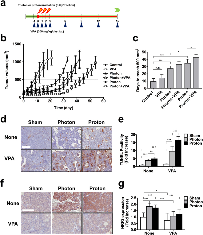
Although efficacy of combined histone deacetylase (HDAC) inhibitors and conventional photon radiotherapy is being tested in clinical trials, their combined effect with proton beam radiotherapy has yet to be determined. Here, we compared combined effect of valproic acid (VPA), a class I and II HDAC inhibitor and antiepileptic drug with proton and photon irradiation in hepatocellular carcinoma (HCC) cells in vitro and in vivo. We found that VPA sensitized more Hep3B cells to proton than to photon irradiation. VPA prolonged proton-induced DNA damage and augmented proton-induced apoptosis. In addition, VPA further increased proton-induced production of intracellular reactive oxygen species and suppressed expression of nuclear factor erythroid-2-related factor 2 (NRF2), a key transcription factor regulating antioxidant response. Downregulation of NRF2 by siRNA transfection increased proton-induced apoptotic cell death, supporting NRF2 as a target of VPA in radiosensitization. In Hep3B tumor xenograft models, VPA significantly enhanced proton-induced tumor growth delay with increased apoptosis and decreased NRF2 expression in vivo. Collectively, our study highlights a proton radiosensitizing effect of VPA in HCC cells. As NRF2 is an emerging prognostic marker contributing to radioresistance in HCC, targeting NRF2 pathway may impact clinical outcome of proton beam radiotherapy.


Conflict of interest statement

The authors declare that they have no competing interests.

Figures

Figure 1
Figure 1
Valproic acid (VPA) inhibits histone deacetylase activity in human hepatocellular carcinoma cells. (a) Western blot analysis shows time-dependent increase of histone H4 acetylation after 1 mM VPA treatment in Hep3B cells. (b) VPA increased histone H4 acetylation in concentration-dependent manner. Samples were harvested 72 h after treatment of indicated concentrations of VPA in Hep3B cells. (c) VPA treatment leads to a modest decrease in proliferation of human hepatocarcinoma Huh7 and Hep3B cells. After 48 h of treatment of indicated concentrations of VPA, cell proliferation was determined using MTT assay. Data are mean values ± SD of 8 samples. *p < 0.05, ***p < 0.001. The cropped blots are presented and their full-length blots are included in the Supplementary Fig. S1.
Figure 2
Figure 2
VPA sensitizes Hep3B cells to proton and photon irradiations. (a) Percentage depth dose graphs in water for photon (solid line) and proton beams (dashed line) indicate different energy distributions. Each arrowhead points to the positions at which cell plates were placed for photon or proton irradiation. (b and c) Clonogenic assay was performed to compare radiation sensitivity. Cells were seeded and irradiated with indicated doses of photon or proton beam with or without 1 mM VPA. After 15 days, survived colonies (>50 cells) were stained and counted. Representative dose-response curves are presented. Data are mean values ± SD of three samples. (b) Proton vs photon; (c) Proton + VPA vs photon + VPA.
Figure 3
Figure 3
VPA attenuates radiation-induced G2/M arrest in Hep3B cells. (a) DNA histogram plots of Hep3B treated with or without 1 mM VPA at 24 h and 72 h after 6 Gy proton or photon irradiation. Cell cycle was assessed by propidium iodide staining and flow cytometry. (b) Distribution of each cell cycle shown as stacked column indicated attenuation of radiation-induced G2/M arrest by VPA. *p < 0.05, **p < 0.01, ***p < 0.001.
Figure 4
Figure 4
VPA attenuates radiation-induced DNA damage repair in Hep3B cells. (a) DNA damage repair after photon or proton irradiation was assessed by γ-H2AX immunostaining. γ-H2AX foci (green) in nuclei (blue) were visualized at 2 h and 24 h after irradiations. Scale bar, 20 µm. (b) Quantification of the number of γ-H2AX foci per cell. Data are mean values ± SD of twelve cells. n.s., not significant; *p < 0.05; **p < 0.01. (c) Western blot analysis reveals combined treatment with proton and VPA led to delayed abrogation of γ-H2AX. (d) Combined treatment with proton and VPA led to persistent activation of ATM and ATR. The samples were harvested 72 h after 6 Gy irradiation. β-actin is used as a loading control. The cropped blots are presented and their full-length blots are included in the Supplementary Fig. S1.
Figure 5
Figure 5
VPA enhances proton-induced apoptosis in Hep3B cells. (a) Population of apoptotic cells at 72 h after radiation treatment was assessed by flow cytometry using Annexin-V staining. Data are mean values ± SD of three samples **p < 0.01. (b) Western blot analysis of cleaved PARP and caspase-3 antibodies showed VPA augmented proton-induced apoptosis. The cropped blots are presented and their full-length blots are included in the Supplementary Fig. S1.
Figure 6
Figure 6
VPA enhances proton-induced ROS production and suppresses activation of NRF2 signaling in Hep3B cells. (a) Production of ROS in 6 Gy photon or proton-irradiated cells was measured using H2DCADA fluorescence dye. Data are mean values ± SD of three samples. *p < 0.05, **p < 0.01, ***p < 0.001. (b) Western blot analysis showed VPA suppressed proton-mediated upregulation of NRF-2 and its downstream HO-1. (c) Western blot analysis confirmed shRNA-mediated depletion of NRF2 in Hep3B. GFP shRNA was used as a control shRNA. (d) Apoptosis assay using flow cytometry with annexin V revealed proton irradiation induced more apoptosis of NRF2-depleted cells than control cells. Data are mean values ± SD of three samples. *p < 0.05, **p < 0.01, ***p < 0.001. The cropped blots are presented and their full-length blots are included in the Supplementary Fig. S1.
Figure 7
Figure 7
VPA enhances proton-induced tumor growth delay in a Hep3B xenograft model. (a) Schematic diagram of the experimental procedure. (b) Administration of VPA further suppressed growth of proton-irradiated tumors. Hep3B cells were implanted into right legs of BALB/c nude mice. Once tumors were palpable, they were irradiated with 3 Gy for 3 consecutive days for a total 9 Gy. Mice were treated with intraperitoneal injections of VPA (300 mg/kg/day) every 3 days. Shown are mean tumor volumes and standard deviation per group (n = 4). (c) Tumour growth delay was determined by calculating days each tumour taken to reach 500 mm3. n.s. not significant; *p < 0.05; ***p < 0.001. (d) TUNEL assay detected more apoptotic cells in tissues co-treated with proton and VPA compared to proton alone or combined photon and VPA. Little was seen in tissues treated with VPA alone. Scale bar, 400 µm. (e) Quantification of the TUNEL positive cell density in tumor tissue sections. n.s. not significant; **p < 0.01; ***p < 0.001. (f) Proton and photon irradiations increased NRF2 expression on tumour tissues, which was suppressed by co-treatment with VPA. NRF2 expression was assessed by immunohistochemistry. Scale bar, 400 µm. (g) Quantification of NRF2 expression in tissue samples. *p < 0.05; ***p < 0.001.

References

    1. Wagner JM, Hackanson B, Lubbert M, Jung M. Histone deacetylase (HDAC) inhibitors in recent clinical trials for cancer therapy. Clin Epigenetics. 2010;1:117–136. doi: 10.1007/s13148-010-0012-4. - DOI - PMC - PubMed
    1. Gray SG, Ekstrom TJ. The human histone deacetylase family. Exp Cell Res. 2001;262:75–83. doi: 10.1006/excr.2000.5080. - DOI - PubMed
    1. Strahl BD, Allis CD. The language of covalent histone modifications. Nature. 2000;403:41–45. doi: 10.1038/47412. - DOI - PubMed
    1. Groselj B, Sharma NL, Hamdy FC, Kerr M, Kiltie AE. Histone deacetylase inhibitors as radiosensitisers: effects on DNA damage signalling and repair. Br J Cancer. 2013;108:748–754. doi: 10.1038/bjc.2013.21. - DOI - PMC - PubMed
    1. West AC, Johnstone RW. New and emerging HDAC inhibitors for cancer treatment. J Clin Invest. 2014;124:30–39. doi: 10.1172/JCI69738. - DOI - PMC - PubMed

Publication types

MeSH terms