. 2017 Nov 8;7(1):15061.
doi: 10.1038/s41598-017-15422-5.

Brain activation induced by chronic psychosocial stress in mice

Affiliations

Brain activation induced by chronic psychosocial stress in mice

Mikaela A Laine et al. Sci Rep. .

Abstract

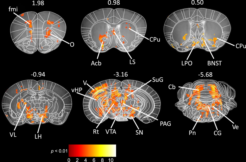
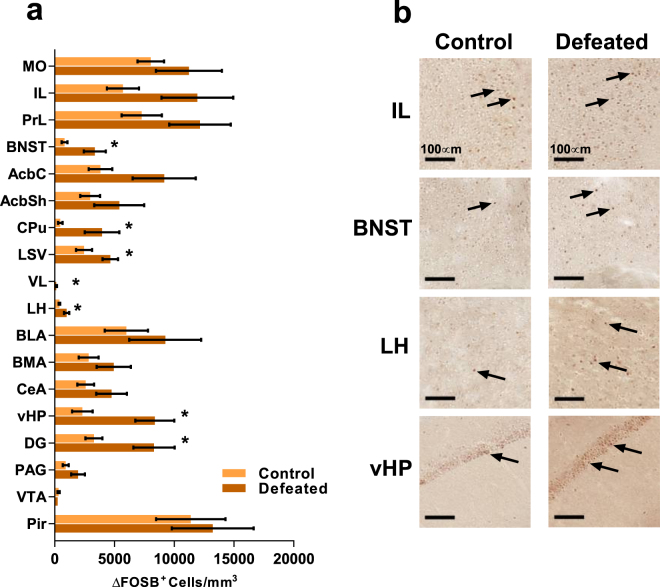
Chronic psychosocial stress is a well-established risk factor for neuropsychiatric diseases. Abnormalities in brain activity have been demonstrated in patients with stress-related disorders. Global brain activation patterns during chronic stress exposure are less well understood but may have strong modifying effects on specific brain circuits and thereby influence development of stress-related pathologies. We determined neural activation induced by chronic social defeat stress, a mouse model of psychosocial stress. To assess chronic activation with an unbiased brain-wide focus we used manganese-enhanced magnetic resonance imaging (MEMRI) and immunohistochemical staining of ∆FOSB, a transcription factor induced by repeated neural activity. One week after 10-day social defeat we observed significantly more activation in several brain regions known to regulate depressive and anxiety-like behaviour, including the prefrontal cortex, bed nucleus of stria terminalis, ventral hippocampus and periaqueductal grey in stressed compared to control mice. We further established that the correlation of ∆FOSB positive cells between specific brain regions was altered following chronic social defeat. Chronic activation of these neural circuits may relate to persistent brain activity changes occurring during chronic psychosocial stress exposure, with potential relevance for the development of anxiety and depression in humans.


Conflict of interest statement

The authors declare that they have no competing interests.

Figures

Figure 1
Figure 1
Timeline of the experiment and illustration of the chronic social defeat stress (CSDS) protocol. (a) Timeline of CSDS, manganese-enhanced magnetic resonance imaging and tissue collection. Each perpendicular line indicates one day. Estimated timeline for detection of Mn2+ by MEMRI and ∆FOSB by immunohistochemical staining is indicated by red and blue lines, respectively. (b) The experimental mouse in the defeated group (dark grey) was placed into the cage of a resident aggressor mouse (white) for a maximum of 10 minutes. It was then placed into the adjacent compartment of the same cage for 24 hours, retaining sensory contact without physical contact. The procedure was repeated for 10 days, each day in a novel resident aggressor mouse’s cage. (c) The control mouse was placed in a compartment adjacent to another control mouse, allowing sensory but not physical contact, for 24 hours. Each day the control mouse was placed into another cage with a novel control mouse, and the procedure was repeated for 10 days. MRI = magnetic resonance imaging.
Figure 2
Figure 2
Differences in brain activity between socially defeated and control mice revealed by MEMRI. Statistical t-maps (thresholded at p < 0.01, corrected for multiple comparisons) are superimposed on a custom-made mouse brain template, with corresponding atlas sections manually overlaid using anatomical landmarks. Numbers indicate the positions of the sections from bregma in millimetres. Brain region abbreviations: BNST (bed nucleus of the stria terminalis), CPu (caudate putamen), Cb (cerebellum), CG (central grey), fmi (forceps minor), LH (lateral hypothalamus), LPO (lateral preoptic area), LS (lateral septum), Acb (nucleus accumbens), O (orbital cortex), PAG (periaqueductal grey), Pn (pontine nuclei), Rt (reticular formation), SN (substantia nigra), SuG (superior colliculus), V (visual cortex), VL (ventrolateral thalamic nucleus), vHP (ventral hippocampus), Ve (vestibular nuclei), VTA (ventral tegmental area).
Figure 3
Figure 3
Chronic social defeat induces changes in brain activation as measured by expression of ∆FOSB. (a) The mean number of cells staining positive for ∆FOSB in defeated and control mice. Error bars ± 1 SEM, *p < 0.05, see Table 1 for exact statistical values. (b) Representative images of immunohistochemical staining for ∆FOSB, positive cells indicated by black arrows, in the infralimbic cortex (IL), bed nucleus of stria terminals (BNST), lateral hypothalamus (LH) and ventral hippocampus (vHP). Scale bar = 100 µm. See Supplementary Fig. S2 for representative images from each of the brain regions analysed.
Figure 4
Figure 4
Chronic social defeat stress associates with alterations in ∆FOSB positive cell number correlation profiles of specific brain regions. Radar graphs depict the absolute value of r for the infralimbic cortex (IL, a), prelimbic cortex (PrL, b), bed nucleus of stria terminalis (BNST, c), basomedial amygdala (BMA, d), ventral hippocampus (vHP, e), and periaqueductal grey (PAG, f) comparison of the number of ∆FOSB-positive cells per mm3 with the other brain regions. Figure key indicates the sign and magnitude of r. Regions that correlate significantly with the title region are indicated by an asterisk (*p < 0.05, **p < 0.01), and correlations remaining significant after correction for false discovery rate (<5%) are marked with bold text. See Table 1 for brain region abbreviations.

References

    1. Kemeny ME, Schedlowski M. Understanding the interaction between psychosocial stress and immune-related diseases: A stepwise progression. Brain Behav Immun. 2007;21:1009–18. doi: 10.1016/j.bbi.2007.07.010. - DOI - PubMed
    1. O’Donnell MJ, et al. Risk factors for ischaemic and intracerebral haemorrhagic stroke in 22 countries (the INTERSTROKE study): A case-control study. The Lancet. 2010;376:112–23. doi: 10.1016/S0140-6736(10)60834-3. - DOI - PubMed
    1. Donner J, et al. Support for involvement of glutamate decarboxylase 1 and neuropeptide y in anxiety susceptibility. Am J Med Genet B Neuropsychiatr Genet. 2012;159B:316–327. doi: 10.1002/ajmg.b.32029. - DOI - PubMed
    1. Moffitt TE, et al. Generalized anxiety disorder and depression: Childhood risk factors in a birth cohort followed to age 32. Psychol Med. 2007;37:441–52. doi: 10.1017/S0033291706009640. - DOI - PubMed
    1. Bick J, Nelson CA. Early adverse experiences and the developing brain. Neuropsychopharmacology. 2016;41:177–196. doi: 10.1038/npp.2015.252. - DOI - PMC - PubMed

Publication types

MeSH terms