Review
. 2017 Oct 25:8:837.
doi: 10.3389/fphys.2017.00837. eCollection 2017.

The Origins and Functions of Tissue-Resident Macrophages in Kidney Development

Affiliations
Review

The Origins and Functions of Tissue-Resident Macrophages in Kidney Development

David A D Munro et al. Front Physiol. .

Abstract

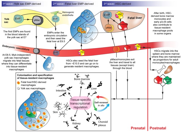
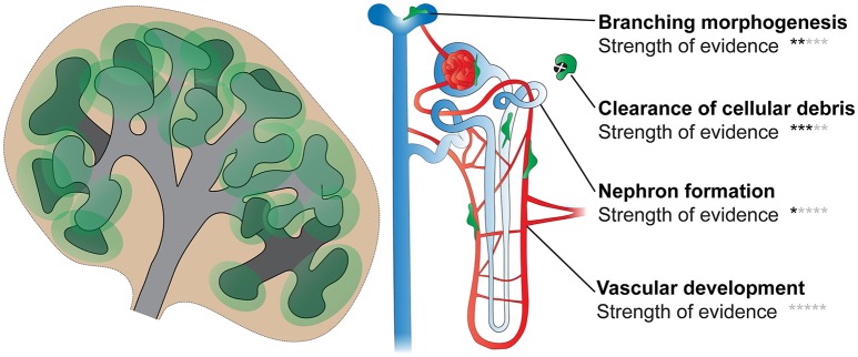
The adult kidney hosts tissue-resident macrophages that can cause, prevent, and/or repair renal damage. Most of these macrophages derive from embryonic progenitors that colonize the kidney during its development and proliferate in situ throughout adulthood. Although the precise origins of kidney macrophages remain controversial, recent studies have revealed that embryonic macrophage progenitors initially migrate from the yolk sac, and later from the fetal liver, into the developing kidney. Once in the kidney, tissue-specific transcriptional regulators specify macrophage progenitors into dedicated kidney macrophages. Studies suggest that kidney macrophages facilitate many processes during renal organogenesis, such as branching morphogenesis and the clearance of cellular debris; however, little is known about how the origins and specification of kidney macrophages dictate their function. Here, we review significant new findings about the origins, specification, and developmental functions of kidney macrophages.

Keywords: angiogenesis; branching morphogenesis; metanephros; monocyte; nephron; ontogeny; phagocyte; renal.


Figures

Figure 1
Figure 1
Overlapping waves of macrophage progenitor cells colonize the developing embryo. Wave 1: Yolk sac-derived erythro-myeloid progenitors (EMPs) differentiate into pMacs and migrate through the early embryo. Wave 2: EMPs, derived from hemogenic endothelium in the yolk sac, enter the fetal liver. In the fetal liver, EMPs expand and differentiate into pMacs and/or monocytes. pMacs/monocytes then egress from the fetal liver and travel through the vasculature into developing organs. Wave 3: Haematopoietic stem cells (HSCs) from the aorta-gonad-mesonephros region also enter the fetal liver and contribute to resident macrophage populations. Some HSCs migrate into the bone marrow and the spleen where they are maintained before being released into the bloodstream postnatally as circulating monocytes that can contribute to tissue-resident macrophage populations. Early pro-B cells are also released from the bone marrow in adulthood and can contribute to certain tissue-resident macrophage populations. Depending on where they engraft, macrophage progenitors start to express transcriptional regulators that define their genetic programme in a tissue-specific manner. EMP, erythro-myeloid progenitor; HSC, haematopoietic stem cell; pMac, pre-macrophage; Camera lucida drawings adapted, with permission, from illustrations by Perry (Gordon et al., 1988).
Figure 2
Figure 2
Contribution of yolk sac-derived and monocyte-derived macrophages to the embryonic and postnatal kidney. The trends shown in the yolk sac-derived and monocyte-derived macrophage graphs are based on fate-mapping experiments by Hoeffel et al. (2015). Trends for the origins of brain macrophages (microglia), which are yolk sac-derived, are shown as a comparison. Adapted with permission from Hoeffel et al. (2015).
Figure 3
Figure 3
Multiple origins of kidney macrophages. Murine kidneys contain macrophages that are derived from multiple sources, with their relative proportions fluctuating throughout development and adulthood. Based on the niche competition hypothesis of macrophage origins (Guilliams and Scott, 2017), we argue that the mixed ontogeny of kidney macrophages is the result of kidney niches being both accessible and available to macrophage precursors throughout kidney development and phases of adulthood. The “?” in some of the yellow bone marrow-derived monocytes denotes that we do not know whether these cells are maintained in the kidney after the resolution of inflammation/disease. The increasing volumes of the cups represent the increasing capacity of the kidney to house macrophages.
Figure 4
Figure 4
Kidney macrophage specification. During development, the tissue-specific signals that kidney macrophages are exposed to provoke the expression of a unique array of transcriptional regulators. Compared to other tissue-resident macrophages, kidney macrophages have increased expression of transcriptional regulators such as Nfatc1, Nfatc2, Ahr, and Irf9. In adulthood, macrophages are exposed to various exogenous stress signals because of factors such as disease, diet, and infection. Based on the multidimensional model of macrophage activation, a macrophage is specified by the integrated effects of the endogenous and exogenous signals within its micro-anatomical site. The macrophage colors are used to show the heterogeneity of macrophage activation status in response to endogenous and exogenous signals in the kidney. DAMPS, damage-associated molecular pattern molecules; PAMPS, pathogen-associated molecular pattern molecules.
Figure 5
Figure 5
Functions of macrophages in kidney development. Processes including branching morphogenesis, cell proliferation/death (and clearance of cellular debris), nephron formation, and vascular development (blood and lymphatic) are important in renal development. Few studies have directly investigated the roles of kidney macrophages in these processes; however, based on available evidence, it is likely that macrophages will facilitate most, if not all, of these processes. Black stars indicate the strength of evidence that macrophages function in each process in kidney development (0 stars, no direct evidence; 5 stars, very strong evidence).

References

    1. Abrahamson D. R. (2009). Development of kidney glomerular endothelial cells and their role in basement membrane assembly. Organogenesis 5, 275–287. 10.4161/org.0.7577 - DOI - PMC - PubMed
    1. Abrahamson D. R., Leardkamolkarn V. (1991). Development of kidney tubular basement membranes. Kidney Int. 39, 382–393. 10.1038/ki.1991.50 - DOI - PubMed
    1. Gonzalez N., Hidalgo A. (2014). Nuclear receptors and clearance of apoptotic cells: stimulating the macrophage's appetite. Front. Immunol. 5:211 10.3389/fimmu.2014.00211 - DOI - PMC - PubMed
    1. Ajami B., Bennett J. L., Krieger C., Tetzlaff W., Rossi F. M. (2007). Local self-renewal can sustain CNS microglia maintenance and function throughout adult life. Nat. Neurosci. 10, 1538–1543. 10.1038/nn2014 - DOI - PubMed
    1. Alikhan M. A., Jones C. V., Williams T. M., Beckhouse A. G., Fletcher A. L., Kett M. M., et al. . (2011). Colony-stimulating factor-1 promotes kidney growth and repair via alteration of macrophage responses. Am. J. Pathol. 179, 1243–1256. 10.1016/j.ajpath.2011.05.037 - DOI - PMC - PubMed

LinkOut - more resources