Review
. 2017:2017:8459402.
doi: 10.1155/2017/8459402. Epub 2017 Oct 8.

Redox Regulation of Inflammatory Processes Is Enzymatically Controlled

Affiliations
Review

Redox Regulation of Inflammatory Processes Is Enzymatically Controlled

Inken Lorenzen et al. Oxid Med Cell Longev. 2017.

Abstract

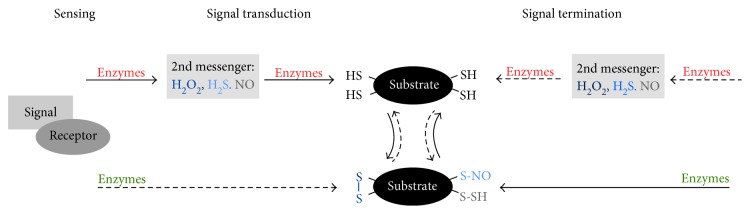
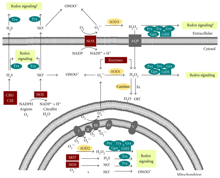
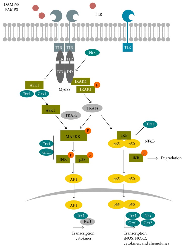
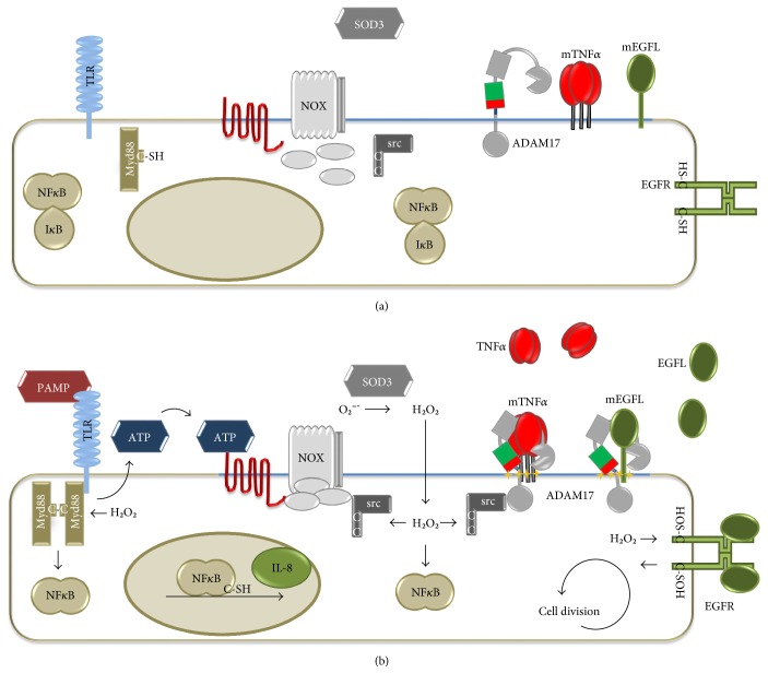
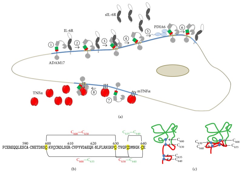
Redox regulation depends on the enzymatically controlled production and decay of redox active molecules. NADPH oxidases, superoxide dismutases, nitric oxide synthases, and others produce the redox active molecules superoxide, hydrogen peroxide, nitric oxide, and hydrogen sulfide. These react with target proteins inducing spatiotemporal modifications of cysteine residues within different signaling cascades. Thioredoxin family proteins are key regulators of the redox state of proteins. They regulate the formation and removal of oxidative modifications by specific thiol reduction and oxidation. All of these redox enzymes affect inflammatory processes and the innate and adaptive immune response. Interestingly, this regulation involves different mechanisms in different biological compartments and specialized cell types. The localization and activity of distinct proteins including, for instance, the transcription factor NFκB and the immune mediator HMGB1 are redox-regulated. The transmembrane protein ADAM17 releases proinflammatory mediators, such as TNFα, and is itself regulated by a thiol switch. Moreover, extracellular redox enzymes were shown to modulate the activity and migration behavior of various types of immune cells by acting as cytokines and/or chemokines. Within this review article, we will address the concept of redox signaling and the functions of both redox enzymes and redox active molecules in innate and adaptive immune responses.


Figures

Figure 1
Figure 1
Concept of redox signaling. A signal is sensed by its receptor, inducing the enzymatic catalyzed production and release of second messengers (e.g., H2O2, NO, and H2S). These activate a cascade of transducing proteins via specific oxidative modifications at Cys residues (e.g., disulfide formation, nitrosylation, and sulfhydration). The effector molecule induces the biological response. A signal can also induce the reduction of distinct Cys residues. The activated signaling cascade becomes terminated, and cysteinyl modifications are reversed. The involved thiol groups are known as thiol switches. Their reduction (green), as well as their oxidation (red) are regulated by different enzymes.
Figure 2
Figure 2
Redox regulation is enzymatically controlled. Illustration of cellular and extracellular enzymes that (i) generate redox active species (red), (ii) decompose reactive species, and are classified as antioxidants (yellow) or (iii) participate in redox signaling (blue). In the cytosol, superoxide (O2) and hydrogen peroxide (H2O2) can be produced by specific enzymes; the cytosolic SOD1 can convert O2 to H2O2. Moreover, the NADPH and oxygen-dependent membrane protein NADPH-oxidase (NOX) can produce O2 that is converted to H2O2 by extracellular SOD3. The latter can cross the membrane via simple diffusion and aquaporins. H2O2 can participate in cell signaling as a second messenger via the action of the thioredoxin family members peroxiredoxin (Prx), thioredoxin (Trx), glutaredoxin (Grx), and glutathione peroxidases. These enzymes are NADPH- and mostly glutathione- (GSH-) dependent. H2O2 can also be reduced to water by the peroxidase catalase, which is mainly located in peroxisomes. However, in the presence of free iron, the highly reactive and damaging hydroxyl radical (OH) is formed from H2O2 via the Fenton reaction. Nitric oxide (NO) is generated by cytosolic NO-synthase (NOS) and hydrogen sulfite (H2S) by the enzymes cystathionine β-synthase (CBS) and cystathionine γ-lyase (CSE). Both constitute second messengers that can participate in redox signaling via the action of Trx. Note that peroxynitrite (ONOO) can spontaneously form in the presence of O2 and NO, inducing irreversible modifications of various biomolecules and thus not participating in redox signaling. In mitochondria, complexes I and III of the mitochondrial respiratory chain produce superoxide (O2−•). Superoxide dismutase 2 (SOD2) converts O2 to H2O2. Mitochondrial NOS and 3-mercaptopyruvate sulfurtransferase (MST) produce NO and H2S, respectively. Mitochondrial H2O2, NO, and H2S can participate in redox signaling. Similar to the cytosol, ONOO and OH can also be formed in the mitochondria. In the extracellular environment, NOX and SOD3 produce O2 and H2O2 and the intracellularly produced NO and H2S can cross the plasma membrane. Members of the Trx family of proteins are found extracellular. Therefore, the intracellular concept of redox signaling might also occur in the microenvironment of the cell.
Figure 3
Figure 3
TLR signaling is redox-regulated. The general concept of the TLR signaling is illustrated, emphasizing the redox-regulated steps and molecules; note that this illustration is simplified and that specific TLR pathways include different proteins. PAMPs and DAMPs are recognized by their specific TLR, which can lead to homo- and heterodimerisation. Upon ligand binding, the TLR associates with the adaptor protein Myd88, which is sensitive to oxidation by hydrogen peroxide and can be regulated by Nrx. Myd88 recruits IRAK4 that phosphorylates IRAK1, which in turn activates additional proteins (e.g., TRAFs and IKK, not shown). MAP kinases and NFκB are activated. MAP kinase signaling is regulated by Trx1 and Grx1 and eventually activates the transcription factor AP1, which has two Cys residues in its DNA binding domain that are reduced by Trx1 via Ref1. The NFκB subunits p50 and p60 are kept in an inhibitory iκB/NFκB-complex in the cytosol. Reduced Trx1 inhibits the dissociation of this complex. Upon dissociation, iκB is phosphorylated and degraded by the proteasome. NF-κB translocates into the nucleus, where it binds to the DNA, a process that depends on the reduction of Cys62 and is regulated by Trx1, Grx1, and/or Nrx. An additional redox-regulated pathway involving ASK1 exists in TLR4 signaling. ASK1 is kept in an inactive complex by reduced Trx1. Upon TLR activation, Trx1 is oxidized, the complex dissociates and active ASK1 regulates JNK activity via different proteins including TRAFs.
Figure 4
Figure 4
Pathogen detection and ROS-dependent defence and regeneration mechanisms. Epithelial cells are constantly exposed to pathogens. The redox state, the localisation, and the activity of different molecules and proteins are altered in the absence (a) or in the presence (b) of pathogens. Activation of TLRs by PAMPs and Myd88 recruitment induce secretion of ATP, which functions as danger signal and activates NOX. TLR and NOX activation both result in NFκB activation, via Myd88 or src, respectively. NFκB translocates to the nucleus and induces the expression of, for example, chemokines such as IL-8, promoting leukocyte recruitment. Myd88 dimerizes upon H2O2 exposure forming disulfide bridges. Src oxidation stabilizes the active conformation of the protease and the oxidation of cysteine residues near the ATP-binding site of the EGFR enhances its activity. Extracellular ATP leads to the activation of the shedding activity of ADAM17. ADAM17 releases soluble TNFα and ligands of the EGFR, such as TGFα and HB-EGF, from the cell surface, whereas TNFα promotes inflammation; signaling via the EGFR leads to regeneration due to induction of cell growth and division (mTNFα: membrane-bound TNFα; mEGFRL: membrane-bound EGFR ligands).
Figure 5
Figure 5
Thiol switch in ADAM17. (a) (1) ADAM17 is active within lipid rafts (blue line). (2) Different stimuli induce the exposure of phosphatidylserine (yellow stars), that interacts with the open and active conformation of the MPD. (3) This process allows ADAM17 to bind and (4) release substrates from the cell surface, for example, soluble interleukin 6 (sIL-6R). (5) Reduced extracellular protein disulfide isomerase PDIA6 catalyzes the disulfide isomerisation targeting the open MPD. (6) The resulting close and inactive structure of ADAM17 is not able to bind and process its substrates. (7) Membrane bound TNFα (mTNFα) is another substrate of ADAM17, (8) which is released upon activation of ADAM17 and also promotes immune response and inflammation. (b) Primary structure of the MPD of human ADAM17, indicating the disulfide bridges involved in the thiol switch. The linear pattern (C600–C630, C635–C640) constitutes the active, the overlaying pattern (C600–C635, C630–C640), the inactive conformation. (c) Structural consequence of the thiol switch of ADAM17. The red-colored part is highly flexible in the open MPD and therefore not visible in the NMR data. The right structure represents the closed conformation of ADAM17 solved by NMR, in which the red part is packed tightly to the upper, green colored part of the MPD.

References

    1. Hanschmann E.-M., Godoy J. R., Berndt C., Hudemann C., Lillig C. H. Thioredoxins, glutaredoxins, and peroxiredoxins—molecular mechanisms and health significance: from cofactors to antioxidants to redox signaling. Antioxidants & Redox Signaling. 2013;19(13):1539–1605. doi: 10.1089/ars.2012.4599. - DOI - PMC - PubMed
    1. Leichert L. I., Dick T. P. Incidence and physiological relevance of protein thiol switches. Biological Chemistry. 2015;396(5):389–399. doi: 10.1515/hsz-2014-0314. - DOI - PubMed
    1. Klotz L.-O., Schroeder P., Sies H. Peroxynitrite signaling: receptor tyrosine kinases and activation of stress-responsive pathways. Free Radical Biology & Medicine. 2002;33(6):737–743. doi: 10.1016/S0891-5849(02)00892-4. - DOI - PubMed
    1. Calcerrada P., Peluffo G., Radi R. Nitric oxide-derived oxidants with a focus on peroxynitrite: molecular targets, cellular responses and therapeutic implications. Current Pharmaceutical Design. 2011;17(35):3905–3932. doi: 10.2174/138161211798357719. - DOI - PubMed
    1. Berndt C., Lillig C. H., Flohé L. Redox regulation by glutathione needs enzymes. Frontiers in Pharmacology. 2014;5:p. 168. doi: 10.3389/fphar.2014.00168. - DOI - PMC - PubMed

LinkOut - more resources