

miR-25 is upregulated before the occurrence of esophageal squamous cell carcinoma
- PMID: 29118908
- PMCID: PMC5666055
miR-25 is upregulated before the occurrence of esophageal squamous cell carcinoma
Abstract
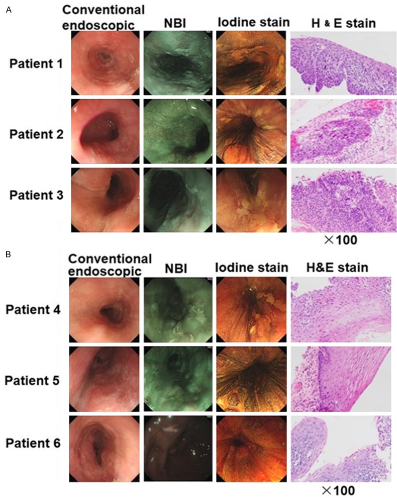
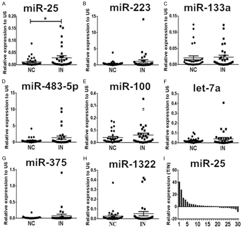
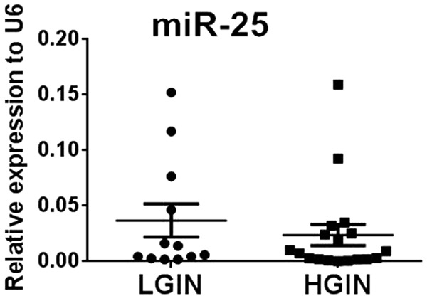
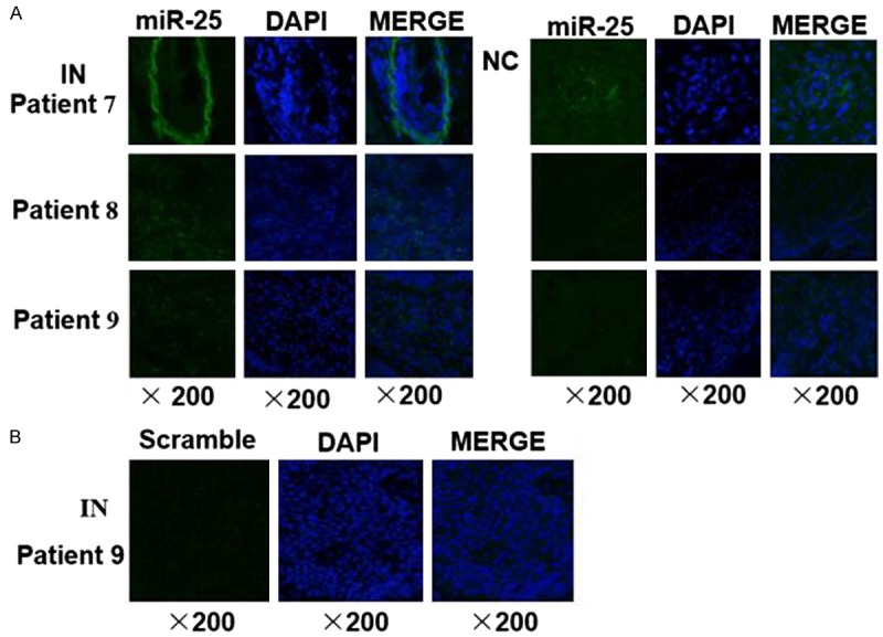
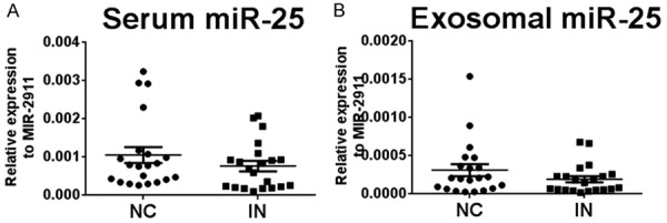
MicroRNAs (miRNAs) are potential biomarkers for cancer detection including esophageal squamous cell carcinoma (ESCC); however, little is known about their expression profile and diagnostic impact in esophageal squamous cell intraepithelial neoplasia, the pathological precancerous lesion of ESCC. In this study, we examined the expression levels of eight miRNAs that were reported to be deregulated in ESCC, including miR-25, let-7a, miR-100, miR-133a, miR-223, miR-375, miR-483-5p and miR-1322, in 30 pairs of esophageal squamous cell neoplasia lesion tissues and corresponding adjacent normal tissues using quantitative real-time PCR (qRT-PCR). Differential expression of miRNAs was further examined by in situ hybridization. Furthermore, the deregulated miRNAs were also measured in serum and serum exosome samples of these patients. miR-25, an oncomir that had been reported to be upregulated in ESCC tissues, were found to be overexpressed in esophageal squamous cell intraepithelial neoplasia lesions (66.7%, 20/30) compared to adjacent normal tissues (P < 0.05), while the other seven miRNAs did not show a significant difference between the lesions and controls. The miR-25 signal was stronger in lesion tissues than in normal tissues according to in situ hybridization. The concentrations of miR-25 in both serum and exosome samples of patients were not significantly different from those of healthy individuals. These findings suggested that the overexpression of miR-25 in esophageal squamous cell intraepithelial neoplasia lesions might be a promising early biomarker candidate for the prediction of ESCC.
Keywords: Esophageal squamous cell carcinoma; esophageal squamous cell intraepithelial neoplasia; in situ hybridization; miR-25; miRNA; qRT-PCR.
Conflict of interest statement
None.
Figures
Similar articles
-
Identification of Novel Circulating miRNA Biomarkers for the Diagnosis of Esophageal Squamous Cell Carcinoma and Squamous Dysplasia.Cancer Epidemiol Biomarkers Prev. 2019 Jul;28(7):1212-1220. doi: 10.1158/1055-9965.EPI-18-1199. Epub 2019 Apr 15. Cancer Epidemiol Biomarkers Prev. 2019. PMID: 30988139
-
Upregulated miR-483-5p expression as a prognostic biomarker for esophageal squamous cell carcinoma.Cancer Biomark. 2017;19(2):193-197. doi: 10.3233/CBM-160506. Cancer Biomark. 2017. PMID: 28211800
-
Promoter hypomethylation mediated upregulation of MicroRNA-10b-3p targets FOXO3 to promote the progression of esophageal squamous cell carcinoma (ESCC).J Exp Clin Cancer Res. 2018 Dec 4;37(1):301. doi: 10.1186/s13046-018-0966-1. J Exp Clin Cancer Res. 2018. PMID: 30514328 Free PMC article.
-
MicroRNAs and esophageal squamous cell carcinoma.Digestion. 2010;82(3):138-44. doi: 10.1159/000310918. Epub 2010 Jun 25. Digestion. 2010. PMID: 20588024 Review.
-
The predictive value of microRNAs for pathological response after neoadjuvant treatment in esophageal squamous cell carcinoma: a systematic review.Ann Transl Med. 2021 Mar;9(5):420. doi: 10.21037/atm-20-3000. Ann Transl Med. 2021. PMID: 33842641 Free PMC article. Review.
Cited by
-
miRNA-425-5p enhances diffuse large B cell lymphoma growth by targeting PTEN.Transl Cancer Res. 2021 Nov;10(11):4905-4913. doi: 10.21037/tcr-21-2394. Transl Cancer Res. 2021. PMID: 35116342 Free PMC article.
-
miR-25 mediates metastasis and epithelial-mesenchymal-transition in human esophageal squamous cell carcinoma via regulation of E-cadherin signaling.Bioengineered. 2019 Dec;10(1):679-688. doi: 10.1080/21655979.2019.1687391. Bioengineered. 2019. PMID: 31679450 Free PMC article.
-
Loss of miR-204-5p Promotes Tumor Proliferation, Migration, and Invasion Through Targeting YWHAZ/PI3K/AKT Pathway in Esophageal Squamous Cell Carcinoma.Onco Targets Ther. 2020 May 26;13:4679-4690. doi: 10.2147/OTT.S243215. eCollection 2020. Onco Targets Ther. 2020. Retraction in: Onco Targets Ther. 2023 Sep 29;16:801-802. doi: 10.2147/OTT.S442447. PMID: 32547097 Free PMC article. Retracted.
-
Exosomes in esophageal cancer: function and therapeutic prospects.Med Oncol. 2024 Nov 27;42(1):18. doi: 10.1007/s12032-024-02543-x. Med Oncol. 2024. PMID: 39601925 Review.
-
Construction of a Nine-MicroRNA-Based Signature to Predict the Overall Survival of Esophageal Cancer Patients.Front Genet. 2021 May 19;12:670405. doi: 10.3389/fgene.2021.670405. eCollection 2021. Front Genet. 2021. PMID: 34093662 Free PMC article.
References
-
- Torre LA, Bray F, Siegel RL, Ferlay J, Lortet-Tieulent J, Jemal A. Global cancer statistics, 2012. CA Cancer J Clin. 2015;65:87–108. - PubMed
-
- Ferlay J, Shin HR, Bray F, Forman D, Mathers C, Parkin DM. Estimates of worldwide burden of cancer in 2008: GLOBOCAN 2008. Int J Cancer. 2010;127:2893–2917. - PubMed
-
- Pennathur A, Gibson MK, Jobe BA, Luketich JD. Oesophageal carcinoma. Lancet. 2013;381:400–412. - PubMed
-
- Tran GD, Sun XD, Abnet CC, Fan JH, Dawsey SM, Dong ZW, Mark SD, Qiao YL, Taylor PR. Prospective study of risk factors for esophageal and gastric cancers in the Linxian general population trial cohort in China. Int J Cancer. 2005;113:456–463. - PubMed
-
- Ruol A, Castoro C, Portale G, Cavallin F, Sileni VC, Cagol M, Alfieri R, Corti L, Boso C, Zaninotto G, Peracchia A, Ancona E. Trends in management and prognosis for esophageal cancer surgery: twenty-five years of experience at a single institution. Arch Surg. 2009;144:247–254. - PubMed
LinkOut - more resources
Full Text Sources