. 2017 Oct 15;9(10):4682-4693.
eCollection 2017.

Topical insulin accelerates cutaneous wound healing in insulin-resistant diabetic rats

Affiliations

Topical insulin accelerates cutaneous wound healing in insulin-resistant diabetic rats

Tianyi Yu et al. Am J Transl Res. .

Abstract

Insulin signaling defects could lead to insulin resistance in insulin target organs: typically, in the muscler, liver, and adipose tissue. We have observed that insulin accelerated diabetic wound healing in our previous works; to further elucidate the mechanism, we investigated the expression and activation of insulin and insulin-like growth factor (IGF)-1 signaling, compared insulin sensitivity in skin tissue with that in liver tissue, and also observed the regulation of insulin on inflammatory response of wounds during the healing process. We found lower expression of insulin receptor, phos-AKT, IGF-1 in type II diabetic rat skin compared with that in normal rat skin. However, the level of phos-AKT in diabetic rat skin remarkably increased after systemic insulin injection, whereas no significant change of phos-AKT was observed in liver upon insulin stimulation. In insulin-treated wounds, we detected a significant increase in insulin signaling proteins and growth factor, as well as the phosphorylated insulin receptor substrate-1 and AKT. The increased Glut1 protein level and translocation of Glut1 from cytosol to cell membrane of the basal epidermal cells were also observed after insulin application. Insulin-treated wounds showed advanced infiltration and resolution of macrophages and a change pattern similar to that of inflammatory mediators, including TNF-α and IL-6. Our findings support that insulin is a valid agent for diabetic wound healing because of its effect on ameliorating defective insulin action and regulating inflammation response. Our results indicate the presence of subtle insulin responsiveness in diabetic skin tissue, regardless of the presence of impaired insulin sensitivity, which could be the cellular and molecular mechanism of insulin accelerating diabetic wound healing.

Keywords: Diabetic; insulin; insulin resistance; wound healing.


Conflict of interest statement

None.

Figures

Figure 1
Figure 1
Diabetic skin showed vigorous responsiveness to insulin although impaired insulin sensitivity was noted. After the induction of diabetes of wistar rats, excision wounds (9-mm diameter) were made in control and diabetic rats; full-thickness skin samples were obtained from the back of diabetic and normal rats for analysis. (A) Western blot analysis was performed to detect protein expression of insulin receptor, IGF-1 receptor, IGF-1, Glut1, phos-AKT, phos-ERK, and (B) TNF-α. β-actin was detected as a loading control. (C) Western blot analysis of phos-AKT in the liver and skin of diabetic rats before (-Ins) or after (+Ins; tissue harvested at 15 min) acute insulin injection. A submaximal insulin dose (0.1 U/kg) was used; liver samples were obtained at basal (-Ins) and 15 min after insulin injection. (D) The quantified expression of the proteins were determined using Image J. Data are shown as the mean ± SD. Significant differences between means were determined by analysis of variance followed by unpaired t-test. *p < 0.05, **p < 0.01, n=6. (E) Represented images of immunofluorescence staining of phos-AKT were detected. Original magnification was 200x and scales bar =50 um.
Figure 2
Figure 2
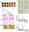
Topical insulin applications improved diabetic wound healing by regulating wound inflammatory cells and repairing cell function. Diabetic wounds were treated with vehicle (20 μL saline solution) or 0.2 U insulin/20 μL saline solution every day until healing was achieved and (A) representative images of the healing process were monitored at day 3, 7, 9, 11, 13, 15 after injury. (B) Wound areas were quantified every other day and expressed as the percentage of wound healing (n=6); statistics are shown as comparisons between the insulin and saline groups. *P < 0.05; **P < 0.01 versus control. (C) Representative H&E stained sections show shorter migrating tongue, fewer vessels in saline wounds at day 7 after wounding, and less mature epidermis after wound healing. Scale bar =1,000 μm. The migration tongues are outlined in red and the reticular edge zone is highlighted in yellow. Blood vessels are indicated by arrows. (D) Immunolabeling for Keratin14, a marker for keratinocyte differentiation, showed that the basal layers of the epidermis of healed wounds are better organized and differentiated when insulin was applied to the wounds. Scales bar =100 μm. (E) Macrophage recruitment on day 3 and 7 after wounding and the day the wound healed is represented by images of immunohistochemical staining for CD68, a specific marker of macrophage. Scale bars=500 μm. Real-time PCR analyzes mRNA expression by comparing fold change of (F) TNF-α and (G) IL-6 mRNA expression at insulin- and saline-treated wounds on day 3 and day 7 after wounding and on the day wound healing was achieved. Data were obtained from 6 wounds, and are shown as mean ± SD. **P < 0.01.
Figure 3
Figure 3
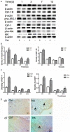
Topical insulin application regulated expression of insulin signaling related proteins on wound area. Diabetic wound were treated with vehicle (20 μL saline solution) or 0.2 U insulin/20 μL saline solution. Wound samples were collected on day 1, 3, 7, and 13 after injury, homogenized, and analyzed. (A) Western blot analysis was performed to detect protein expression of insulin receptor, IGF-1 receptor, phos-IRS-1, IGF-1 and phos-AKT. The antibody dilution ratio is 1:1000. β-actin was detected as a loading control. (B, C) The quantified expression of the proteins were determined using Image J. Data are shown as the mean value ± SD (n=6, *p < 0.05; **p < 0.01 versus control). Significant differences between means were determined by analysis of variance followed by unpaired t-test. (D) Representative immunohistochemical sections showed an increased number of phos-AKT- expressing cells in insulin-treated wounds at day 3 and 7. Scale bar =125 μm.
Figure 4
Figure 4
Topical insulin application regulated expression of Glut1. Diabetic wound samples treated with vehicle (20 μL saline solution) or 0.2 U insulin/20 μL saline solution were collected on day 1, 3, 7, and 13 after injury, homogenized, and analyzed. (A) Glut1 levels were detected by western blot analysis. The antibody dilution ratio is 1:1000. β-actin was detected as a loading control. (B) The quantified expression was determined using Image J. Data are shown as the mean ± SD (n=6, **p < 0.01). Statistics are shown as comparisons between the treatment and control groups. (C) Images immunostained with antibodies specific to Glut1 are presented. Insulin treatment promotes Glut1’s translocation to the plasma membrane, which was found to be increased 30% on day 3 and 60% on day 7 compared with controls.

Similar articles

Cited by

References

    1. Joseph B. Insulin in the treatment of non-diabetic bed sores. Ann Surg. 1930;92:318–319. - PMC - PubMed
    1. Rosenthal SP. Acceleration of primary wound healing by insulin. Arch Surg. 1968;96:53–55. - PubMed
    1. Liu Y, Petreaca M, Yao M, Martins-Green M. Cell and molecular mechanisms of keratinocyte function stimulated by insulin during wound healing. BMC Cell Biol. 2009;10:1. - PMC - PubMed
    1. Liu Y, Petreaca M, Martins-Green M. Cell and molecular mechanisms of insulin-induced angiogenesis. J Cell Mol Med. 2009;13:4492–4504. - PMC - PubMed
    1. Chen X, Liu Y, Zhang X. Topical insulin application improves healing by regulating the wound inflammatory response. Wound Repair Regen. 2012;20:425–434. - PubMed

LinkOut - more resources