. 2017:2017:9182748.
doi: 10.1155/2017/9182748. Epub 2017 Oct 8.

Effects of Propofol Treatment in Neural Progenitors Derived from Human-Induced Pluripotent Stem Cells

Affiliations

Effects of Propofol Treatment in Neural Progenitors Derived from Human-Induced Pluripotent Stem Cells

Bo Long et al. Neural Plast. 2017.

Abstract

Propofol is an intravenous anesthetic that has been widely used in clinics. Besides its anesthetic effects, propofol has also been reported to influence the regulation of the autonomic system. Controversies exist with regard to whether propofol exposure is safe for pregnant women and young children. In this work, human-induced pluripotent stem cell- (hiPSC-) derived neural progenitor cells (NPCs) were treated with propofol at 20, 50, 100, or 300 μM for 6 h or 24 h, and acute and subacute cell injury, cell proliferation, and apoptosis were evaluated. Comparison of genome-wide gene expression profiles was performed for treated and control iPSC-NPCs. Propofol treatment for 6 h at the clinically relevant concentration (20 or 50 μM) did not affect cell viability, apoptosis, or proliferation, while propofol at higher concentration (100 or 300 μM) decreased NPC viability and induced apoptosis. In addition, 20 μM propofol treatment for 6 h did not alter global gene expression. In summary, propofol treatment at commonly practiced clinical doses for 6 h did not have adverse effects on hiPSC-derived NPCs. In contrast, longer exposure and/or higher concentration could decrease NPC viability and induce apoptosis.


Figures

Figure 1
Figure 1
Generation of NPCs from hiPSCs. Representative images of neural tube structures generated from differentiating NES-GFP reporter hiPSC line via embryoid body formation method on day 6. GFP serves as a surrogate marker for NESTIN, a widely accepted NPC marker (a). The neural rosettes were attached to culture plates on day 10 as monolayer culture which continued to express GFP (NESTIN) (b). Similarly, NESTIN and another NPC marker SOX1 were both expressed robustly and uniformly in NPCs that were derived from two additional hiPSC lines, USCK7 (c) and ND2-0 (d), as revealed by immunocytochemistry staining of both NESTIN (green) and SOX1 (red). DAPI (blue) was used to reveal nuclei. Bar, 50 μm.
Figure 2
Figure 2
High-concentration propofol treatment exerts toxicity to NPCs. NPCs derived from three hiPSC lines were exposed to propofol at different concentrations (0, 20, 50, 100, and 300 μM). MTT assays showed high-concentration propofol-reduced NPCs viability after the 6 h (a) or 24 h (c) treatment, or 6 h treatment followed by 20 h washout (b). Data from LDH assays further showed that propofol at a high concentration reduced the number of NPCs (d). Data at each concentration were analyzed by unpaired t-tests. For MTT assays, data were expressed as a percentage of viable cells of the treatment compared to the untreated (0 μM propofol) group (mean ± SD). For LDH assays, data were expressed as a percentage of positive control (mean ± SD), compared to 0 μM propofol exposure group. For both assays, 5 wells per treatment condition were examined. Each experiment was repeated for at least three times. p < 0.05; ∗∗p < 0.01; ∗∗∗p < 0.001.
Figure 3
Figure 3
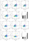
Propofol-induced apoptosis in NPCs. NPCs derived from USCK7 and ND2-0 were treated with propofol at 0, 20, 50, 100, and 300 μM for 6 h and apoptotic cells quantified by flow cytometry of FITC-labelled Annexin V. The percentage of different cell populations is shown in each of the four quadrants of the representative flow cytometry charts, and the statistical analyses are summarized in (USCK7) (f) and (ND2-0) (i). The percentage of apoptotic cells is shown at the left lower quadrant of each chart. For USCK7-NPCs, the 100 and 300 μM propofol treatment groups showed significantly higher percentage of apoptotic cells (a, b, c, d, e, f). For ND2-0 NPCs, only the 300 μM treatment group showed a significantly higher percentage of apoptotic cells (g, h, i, j, k, l). Data were expressed as a percentage of FITC+/DAPI− cells (mean ± SD) for n = 3 flow cytometry experiments per treatment condition. p < 0.05; ∗∗p < 0.01; ∗∗∗p < 0.001.
Figure 4
Figure 4
Propofol treatment for 6 h did not affect NPC proliferation. NPCs derived from three hiPSC lines were treated with propofol at different concentrations (0, 20, 50, 100, and 300 μM). Cell proliferation was assessed by Ki-67 (red) immunocytochemistry staining. Nuclei were revealed by DAPI (blue) (a, b, c). At least 1000 cells were counted for each experiment. Data were expressed as percentage of Ki-67+ cells (mean ± SD). n = 3 Ki-67 staining per treatment condition. Bar, 50 μm.
Figure 5
Figure 5
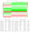
Gene expression profile of propofol-treated NPCs. NES-GFP iPSC-derived NPCs were treated with propofol (20 μM or 300 μM) for 6 h, and RNAs extracted immediately for Illumina BeadArray. Heatmap (a) of hierarchical clustering of differentially expressed genes (DEGs) shows that the 20 μM propofol-treated group clustered together with the untreated group, while the 300 μM propofol-treated group shows distinct gene expression profile. The top ten upregulated and downregulated DEGs are listed in (b).
Figure 6
Figure 6
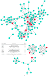
PPI network construction of DEGs extracted from comparison of treated (300 μM) with untreated NPCs. The PPI relationships of the DEGs were obtained by using search tool for the retrieval of interacting genes (STRING) and visualized using the Cytoscape 3 software. After filtering out disconnected DEGs, a network with 101 nodes and 251 edges were obtained with the combined score > 0.4 (a). The connectivity degree of each node of the PPI network was calculated. Eight genes with connectivity degree > 10 were selected as hub genes (b). Clusters with densely connected regions based on topology were built with Molecular COmplex DEtection (MCODE). The top three subnetworks represent important cellular and molecular pathways including aminoacyl-tRNA biosynthesis (c), oxidative phosphorylation (d), and cell cycle (e).
Figure 7
Figure 7
Verification of the expression level of genes related to UPR and ER press. NES-GFP iPSC-derived NPCs were treated with propofol (0, 20, 50, 100, or 300 μM) for 6 h. The mRNA expression level of ATF4, CEBPB, DDIT3, and TRIB3 was evaluated by microarray (a, b, c, d) and qRT-PCR (e, f, g, h). The qRT-PCR results were normalized to the GAPDH mRNA level. All data are presented as mean ± SD (n = 3; p < 0.05; ∗∗∗p < 0.001, Student's t-test).

Similar articles

Cited by

References

    1. Heid F., Kauff D. W., Lang H., Kneist W. Impact of inhalation vs. intravenous anaesthesia on autonomic nerves and internal anal sphincter tone. Acta Anaesthesiologica Scandinavica. 2015;59(9):1119–1125. doi: 10.1111/aas.12535. - DOI - PubMed
    1. Hidaka S., Kawamoto M., Kurita S., Yuge O. Comparison of the effects of propofol and midazolam on the cardiovascular autonomic nervous system during combined spinal and epidural anesthesia. Journal of Clinical Anesthesia. 2005;17(1):36–43. doi: 10.1016/j.jclinane.2004.03.012. - DOI - PubMed
    1. Martini C. H., Boon M., Broens S. J., et al. Ability of the nociception level, a multiparameter composite of autonomic signals, to detect noxious stimuli during propofol-remifentanil anesthesia. Anesthesiology. 2015;123(3):524–534. doi: 10.1097/ALN.0000000000000757. - DOI - PubMed
    1. Chidambaran V., Costandi A., D'Mello A. Propofol: a review of its role in pediatric anesthesia and sedation. CNS Drugs. 2015;29(7):543–563. doi: 10.1007/s40263-015-0259-6. - DOI - PMC - PubMed
    1. Kuczkowski K. M. The safety of anaesthetics in pregnant women. Expert Opinion on Drug Safety. 2006;5(2):251–264. doi: 10.1517/14740338.5.2.251. - DOI - PubMed

MeSH terms