. 2018 Jun;75(11):2011-2026.
doi: 10.1007/s00018-017-2705-8. Epub 2017 Nov 9.

Re-evaluation of protein kinase CK2 pleiotropy: new insights provided by a phosphoproteomics analysis of CK2 knockout cells

Affiliations

Re-evaluation of protein kinase CK2 pleiotropy: new insights provided by a phosphoproteomics analysis of CK2 knockout cells

Cinzia Franchin et al. Cell Mol Life Sci. 2018 Jun.

Abstract

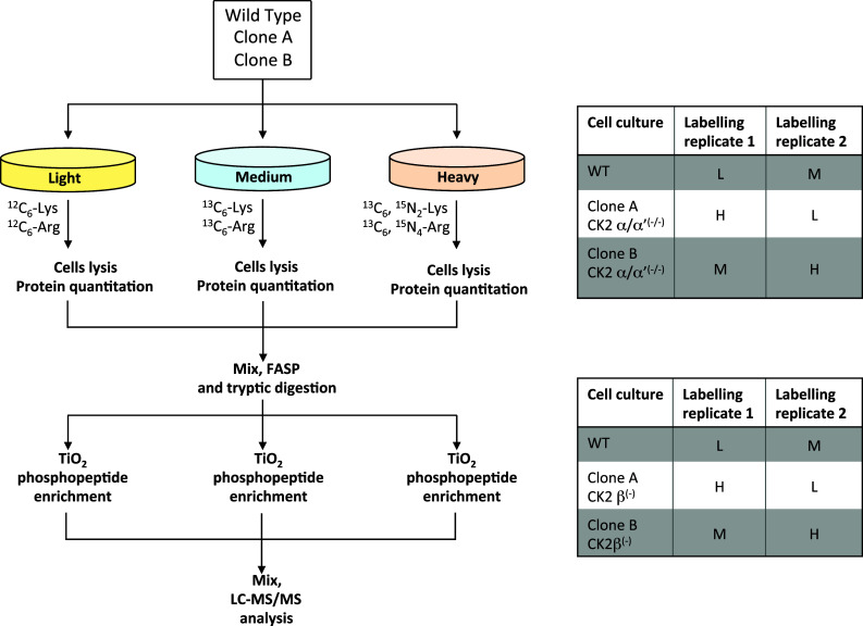
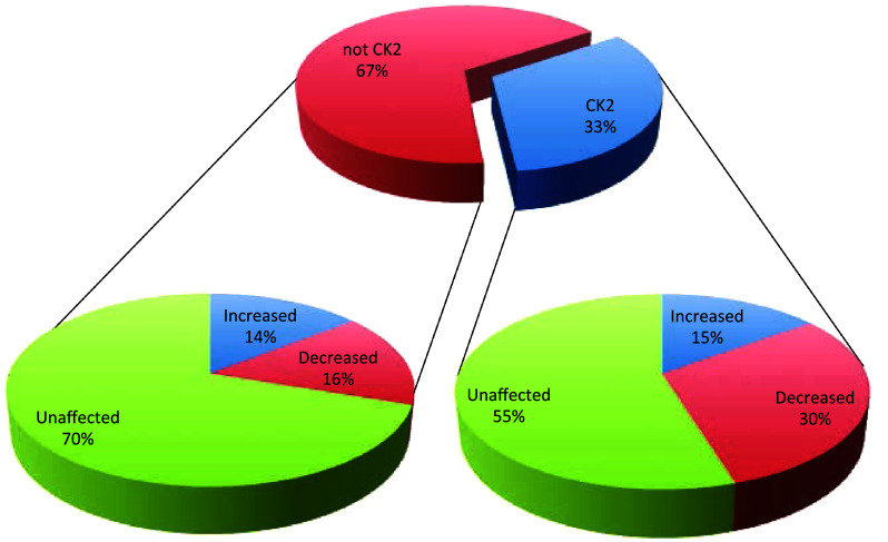
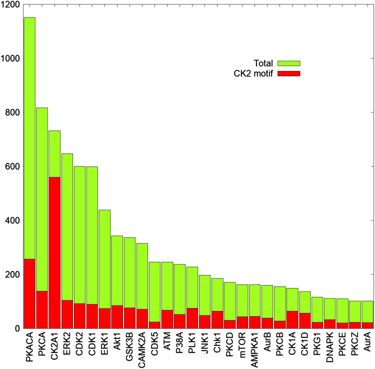
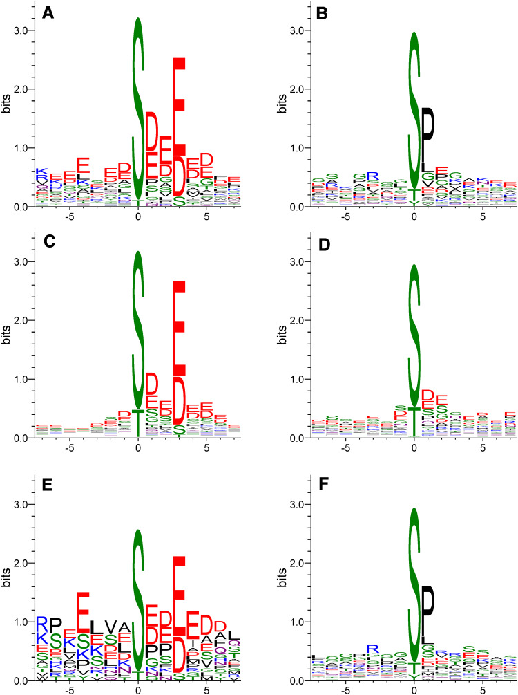
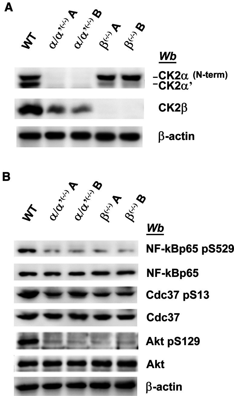
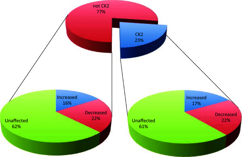
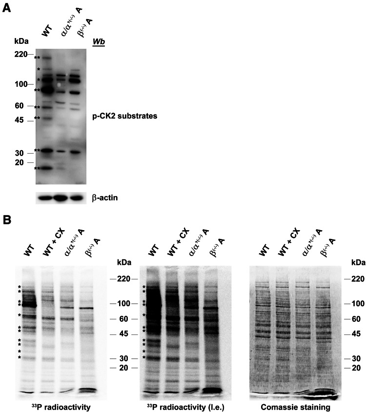
CK2 denotes a ubiquitous and pleiotropic protein kinase whose holoenzyme is composed of two catalytic (α and/or α') and two regulatory β subunits. The CK2 consensus sequence, S/T-x-x-D/E/pS/pT is present in numerous phosphosites, but it is not clear how many of these are really generated by CK2. To gain information about this issue, advantage has been taken of C2C12 cells entirely deprived of both CK2 catalytic subunits by the CRISPR/Cas9 methodology. A comparative SILAC phosphoproteomics analysis reveals that, although about 30% of the quantified phosphosites do conform to the CK2 consensus, only one-third of these are substantially reduced in the CK2α/α'(-/-) cells, consistent with their generation by CK2. A parallel study with C2C12 cells deprived of the regulatory β subunit discloses a role of this subunit in determining CK2 targeting. We also find that phosphosites notoriously generated by CK2 are not fully abrogated in CK2α/α'(-/-) cells, while some phosphosites unrelated to CK2 are significantly altered. Collectively taken our data allow to conclude that the phosphoproteome generated by CK2 is not as ample and rigidly pre-determined as it was believed before. They also show that the lack of CK2 promotes phosphoproteomics perturbations attributable to kinases other than CK2.

Keywords: Cell signalling; Kinase inhibitors; Protein phosphorylation; Quantitative proteomics; Sequence logo.


Conflict of interest statement

The authors declare that they have no conflict of interest.

Figures

Fig. 1
Fig. 1
Workflow of the SILAC experiments. The figure summarises the experimental set-up that was used to compare the phosphoproteomes of C2C12 wild type cells with those relative to CK2α/α′(−/−) cells (clone A and clone B) and CK2β(−/−) cells (clone A and clone B). Two biological replicates with a label-swap strategy were performed for each of the triplex SILAC experiments. To reduce the variability associated to the phosphopeptide enrichment step, the TiO2 affinity purification was performed in triplicate for each biological replicate and phosphopeptides were then pooled before LC–MS/MS analysis
Fig. 2
Fig. 2
Pie charts summarising the results obtained from the SILAC experiment of wild type vs CK2α/α′(−/−) cells. The central pie chart shows the percentage of phosphosites (reliably quantified in the SILAC experiment of wild type vs CK2α/α′(−/−) cells) that either conform (CK2 consensus) or do not conform (not CK2 consensus) to the canonical consensus sequence of the kinase (pS/pT-X-X-E/D/pSpT). For each of these two categories, the percentage of phosphosites that are either significantly increased or decreased, or that do not show significant changes in their abundance are reported in the lateral pie charts
Fig. 3
Fig. 3
CK2 consensus is present in numerous phosphosites generated by the most pleiotropic protein kinases. The graph shows the total number of phosphosites reported in the kinase substrate database of PhosphoSitePlus for each of the most pleiotropic protein kinases. Red bars indicate the number of phosphosites that conform also to the canonical consensus sequence of CK2
Fig. 4
Fig. 4
Sequence logo analysis. Sequence Logos obtained from all phosphopeptides either conforming (panel A) or not conforming (panel B) to the CK2 consensus sequence and characterised by a significantly reduced phosphorylation level in CK2α/α′(−/−) with respect to wild type C2C12 cells. Panel C and D display the Sequence Logos obtained from all phosphosites attributed to CK2 in the PhosphoSitePlus database and that either conform (panel C) or do not conform (panel D) to the canonical consensus sequence of CK2. Sequence Logos obtained from all phosphopeptides either conforming (panel E) or not conforming (panel F) to the CK2 consensus sequence and characterised by a significantly reduced phosphorylation level in CK2β(−/−) with respect to wild type C2C12 cells
Fig. 5
Fig. 5
Western blot analysis of CK2-substrates in wild type and CK2-KO cells. a, b Cellular lysates of wild type (WT), two different CK2α/α′(−/−) clones (A and B), and two different CK2β(−/−) clones (A and B) were analysed by western blot with the antibodies that specifically recognise phosphosites generated by CK2, as indicated. β-actin was used as loading control. Figures are representative of three separate experiments
Fig. 6
Fig. 6
Pie charts summarising the results obtained from the SILAC experiment of wild type vs CK2β(−/−) cells. The central graph shows the percentage of phosphosites (reliably quantified in the SILAC experiment of wild type vs CK2β(−/−) cells) that either conform (CK2 consensus) or do not conform (not CK2 consensus) to the canonical consensus sequence of the kinase. For each of these two categories, the percentage of phosphosites that are either significantly increased or decreased, or that do not show significant changes in their abundance are reported in the lateral pie charts
Fig. 7
Fig. 7
Comparison of global CK2 substrate phosphorylation in CK2α/α′ and in CK2β knockout clones. a Cellular lysates of wild type (WT), CK2α/α′(−/−) and CK2β(−/−) were analysed by western blot with a specific antiphospho-CK2 substrate antibody, as described in the experimental section. β-actin was used as loading control. CK2 substrates that decrease their phosphorylation both in CK2α/α′(−/−) and CK2β(−/−) are denoted by two asterisks, while substrates that display a decreased phosphorylation level only in CK2α/α′(−/−) are denoted by a single asterisk. b In vitro phosphorylation of cell lysates. 15 μg of cell lysates were incubated in the presence of [γ-33P]ATP as described in the Methods section. Where indicated, 500 nM of CX-4945 (CX) was added to the phosphorylation reaction. After 10 min at 37 °C, proteins were separated by electrophoresis on SDS-PAGE, coomassie stained and analysed by PhosphorImager (l.e. means long exposition). Radioactive bands whose intensity decreases in CK2α/α′(−/−) are denoted by an asterisk. Figures are representative of at least four independent experiments

References

    1. Allende CC, Allende JE. Promiscuous subunit interactions: a possible mechanism for the regulation of protein kinase CK2. J Cell Biochem Suppl. 1998;30–31:129–136. doi: 10.1002/(SICI)1097-4644(1998)72:30/31+<129::AID-JCB17>3.0.CO;2-P. - DOI - PubMed
    1. Pinna LA. Protein kinase CK2: a challenge to canons. J Cell Sci. 2002;115(Pt 20):3873–3878. doi: 10.1242/jcs.00074. - DOI - PubMed
    1. St-Denis NA, Litchfield DW. Protein kinase CK2 in health and disease: from birth to death: the role of protein kinase CK2 in the regulation of cell proliferation and survival. Cell Mol Life Sci. 2009;66(11–12):1817–1829. doi: 10.1007/s00018-009-9150-2. - DOI - PMC - PubMed
    1. Pinna LA. The raison d’etre of constitutively active protein kinases: the lesson of CK2. Acc Chem Res. 2003;36(6):378–384. doi: 10.1021/ar020164f. - DOI - PubMed
    1. Litchfield DW. Protein kinase CK2: structure, regulation and role in cellular decisions of life and death. Biochem J. 2003;369(Pt 1):1–15. doi: 10.1042/bj20021469. - DOI - PMC - PubMed

Publication types

LinkOut - more resources