

Review
doi: 10.1007/s00441-017-2706-9.
Epub 2017 Nov 8.
Neurodegeneration and the ordered assembly of α-synuclein
Affiliations
- PMID: 29119326
- PMCID: PMC6015613
- DOI: 10.1007/s00441-017-2706-9
Item in Clipboard
Review
Neurodegeneration and the ordered assembly of α-synuclein
Cell Tissue Res.
2018 Jul.
Abstract
In 2017, it was 200 years since James Parkinson published 'An Essay on the Shaking Palsy' and 20 years since α-synuclein aggregation came to the fore. In 1998, multiple system atrophy joined Parkinson's disease and dementia with Lewy bodies as the third major synucleinopathy. Here, we describe the work that led to the identification of α-synuclein in Lewy bodies, Lewy neurites and Papp-Lantos bodies. We also review some of the findings reported since 1997.
Keywords: Alpha-synuclein; Dementia with Lewy bodies; Multiple system atrophy; Ordered assembly; Parkinson’s disease.
Figures

The α-synuclein pathology of Parkinson’s disease. Lewy pathology in the substantia nigra and several other brain regions defines Parkinson’s disease at the neuropathological level. This is shown by light microscopy, labelled by α-synuclein antibodies (a–c). Using immunoelectron microscopy, filaments extracted from the Lewy pathology were decorated by α-synuclein antibodies (d–g). a Two pigmented nerve cells, each containing an α-synuclein-positive Lewy body (red arrows); Lewy neurites (black arrows) are also immunopositive. Scale bar 20 μm. b Pigmented nerve cell with two α-synuclein-positive Lewy bodies. Scale bar 8 μm. c α-Synuclein-positive extracellular Lewy body. Scale bar 4 μm. d–g Isolated filaments from the substantia nigra of patients with Parkinson’s disease are decorated by an antibody directed against the carboxy-terminal (d, e) or the amino-terminal (f, g) region of α-synuclein. The gold particles conjugated to the second antibody appear as black dots. Note the uniform decoration (d, e) and the labelling of only one filament end (f, g). Scale bar 100 nm. From Goedert (2001)

The α-synuclein pathology of multiple system atrophy. Glial cytoplasmic inclusions in several brain regions define multiple system atrophy. Similar inclusions are also present in the nuclei of some glial cells, as well as in the cytoplasm and nuclei of some nerve cells and in nerve cell processes. Inclusions labelled by α-synuclein antibodies are shown by light microscopy (a–c). Using immunoelectron microscopy, filaments extracted from the inclusions were decorated by α-synuclein antibodies (d–g). a α-Synuclein-immunoreactive cytoplasmic oligodendrocyte inclusions (red arrows) in pontine fibre tracts. b α-Synuclein-immunoreactive nuclear oligodendrocyte inclusion (red arrow) and cytoplasmic nerve cell inclusion (black arrow) in grey matter of frontal cortex. c α-Synuclein-immunoreactive nuclear nerve cell inclusion (black arrow) in grey matter of pons. Scale bars (a) 5 μm, (b) 50 μm, (c) 30 μm. d–g Isolated filaments from the frontal cortex and cerebellum of patients with multiple system atrophy are decorated by antibodies specific for the carboxy-terminal (d, e) and amino-terminal (f, g) regions of α-synuclein. The gold particles conjugated to the second antibody appear as black dots. Note the uniform decoration in (d, e) and the labelling of only one filament end in (f, g). A twisted filament is shown in (d), whereas (e) shows a straight filament. Scale bar 100 nm. Adapted from Goedert (2001)
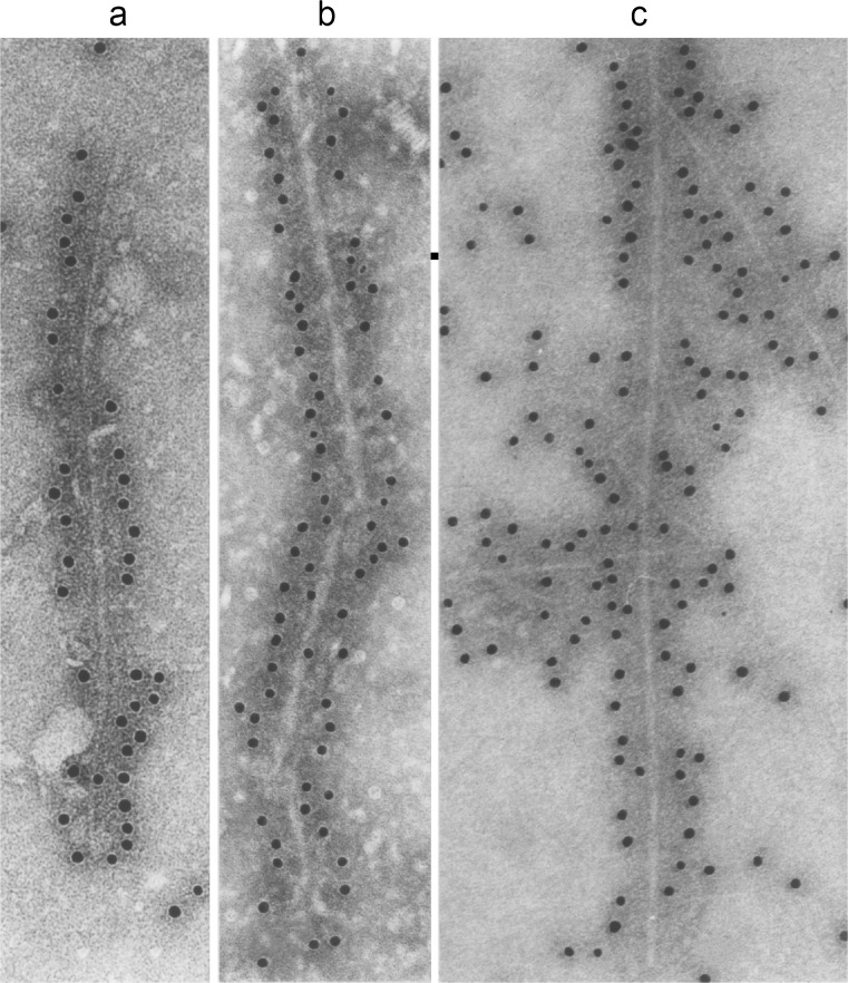
Filaments extracted from the brains of patients with dementia with Lewy bodies (a), multiple system atrophy (b) or assembled from bacterially expressed human α-synuclein (c) were decorated by an anti-α-synuclein antibody. The gold particles conjugated to the second antibody appear as black dots. From Goedert and Spillantini (2012)

Human α-synuclein and its disease-causing mutations. a Diagram of the 140 amino acid human α-synuclein. The seven amino-terminal repeats are shown as blue bars. b A dominantly inherited increase in gene dosage (duplication or triplication) of the chromosomal region containing SNCA gives rise to Parkinson’s disease and dementia with Lewy bodies. Homozygous duplications have also been described. In addition, missense mutations in SNCA cause dominantly inherited forms of Parkinson’s disease and dementia with Lewy bodies. c The repeats (residues 7–87) of human α-synuclein are shown, with disease-causing mutations (A30P, E46K, H50Q, G51D, A53E, A53T and A53V) given as blue letters. Amino acids that are identical in at least five of the seven repeats are shaded in blue
References
-
- Abeliovich A, Schmitz Y, Farinas I, Choi-Lundberg D, Ho WH, Castillo PE, Hinsky N, Verdugo JM, Armanini M, Ryan A, Hynes M, Phillips H, Sulzer D, Rosenthal A. Mice lacking α-synuclein display functional deficits in the nigrostriatal dopamine system. Neuron. 2000;25:239–252. - PubMed
-
- Ansari KA, Johnson AJ. Olfactory function in Parkinson’s disease. J Chronic Dis. 1975;28:493–497. - PubMed
-
- Anwar S, Peters O, Millership S, Ninkina NN, Doig N, Connor-Robson N, Threlfell S, Kooner G, Deacon RM, Bannerman DM, Bolam JP, Chandra SS, Cragg SJ, Wade-Martins R, Buchman VL. Functional alterations to the nigrostriatal system in mice lacking all three members of the synuclein family. J Neurosci. 2011;31:7264–7274. - PMC - PubMed
Publication types
MeSH terms
Substances
Grants and funding
LinkOut - more resources
Full Text Sources
Other Literature Sources
