Review
. 2017 Nov 9:3:17083.
doi: 10.1038/nrdp.2017.83.

Rotavirus infection

Affiliations
Review

Rotavirus infection

Sue E Crawford et al. Nat Rev Dis Primers. .

Abstract

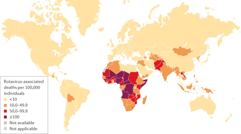
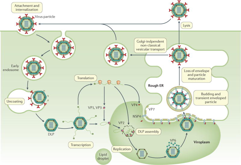
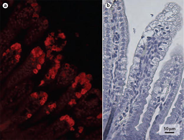
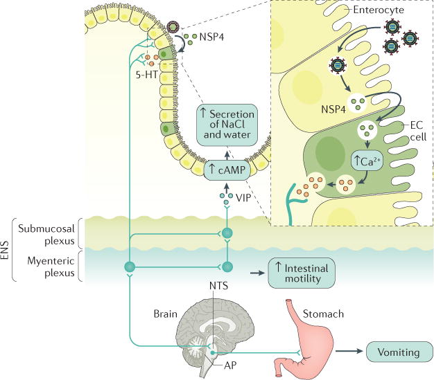
Rotavirus infections are a leading cause of severe, dehydrating gastroenteritis in children <5 years of age. Despite the global introduction of vaccinations for rotavirus over a decade ago, rotavirus infections still result in >200,000 deaths annually, mostly in low-income countries. Rotavirus primarily infects enterocytes and induces diarrhoea through the destruction of absorptive enterocytes (leading to malabsorption), intestinal secretion stimulated by rotavirus non-structural protein 4 and activation of the enteric nervous system. In addition, rotavirus infections can lead to antigenaemia (which is associated with more severe manifestations of acute gastroenteritis) and viraemia, and rotavirus can replicate in systemic sites, although this is limited. Reinfections with rotavirus are common throughout life, although the disease severity is reduced with repeat infections. The immune correlates of protection against rotavirus reinfection and recovery from infection are poorly understood, although rotavirus-specific immunoglobulin A has a role in both aspects. The management of rotavirus infection focuses on the prevention and treatment of dehydration, although the use of antiviral and anti-emetic drugs can be indicated in some cases.


Conflict of interest statement

Competing interests

The authors declare no competing interests.

Figures

Figure 1
Figure 1. Rotavirus structure
The rotavirus particle resembles a wheel with short spokes and a smooth outer rim. a | Electron micrograph of rotavirus triple-layered particles. b | Cross-sectional schematic of the rotavirus triple-layered particle. This structure consists of the inner capsid layer (viral protein (VP)2), the middle capsid layer (VP6) and the outer capsid layer (VP7 and the spike protein VP4). VP4 is proteolytically cleaved into VP8* and VP5*. The structural protein VP2, the enzymes VP1 and VP3 and the viral genome compose the virion core. The middle capsid layer protein (VP6) determines species, group and subgroup specificities. The outer capsid layer is composed of two proteins, VP7 and VP4, which elicit an immune response in infected hosts, leading to the production of rotavirus-specific antibodies. c| Electrophoretic migration profile of the 11 segments of rotavirus double-stranded RNA (dsRNA) and the encoded proteins for simian rotavirus SA11 strain. NSP, non-structural protein.
Figure 2
Figure 2. Rotavirus-associated mortality in children <5 years of age in 2013
As of April 2016, the WHO estimated that worldwide, 215,000 children <5 years of age died because of rotavirus infection in 2013. Figure reproduced with permission from the World Health Organization. Rotavirus mortality rate in children younger than 5 years, 2013. http://www.who.int/immunization/monitoring_surveillance/burden/estimates/rotavirus/rotavirus_deaths_map_b.jpg?ua=1 (2017) (REF. 208).
Figure 3
Figure 3. The number of rotavirus-positive tests in the United States before and after vaccine introduction
These data are from 21 continuously reporting National Respiratory and Enteric Viruses Surveillance System laboratories, collected by week of year and region, including a 3-week moving average. The number of rotavirus-positive tests has decreased, and the annual peak in rotavirus cases has been replaced by a biennial peak, following the introduction of rotavirus vaccinations (dotted line),. Figure adapted with permission from REF. , Centers for Disease Control and Prevention.
Figure 4
Figure 4. The rotavirus replication cycle
Rotaviruses attach to different glycan receptors on the host cell surface, depending on the virus strain, through interaction with the viral protein 8* (VP8*) domain of VP4. For decades, sialoglycans (for example, gangliosides such as GM1 and GD1a) were considered the key cellular glycan partner for VP8* (REF. 37). Although this remains true for animal rotavirus strains, VP8* of many human rotavirus strains binds genetically determined nonsialylated glycoconjugates, called histo-blood group antigens (HBGAs). Other proposed receptors for rotavirus cell entry include integrins, heat shock protein 70 (REFS 211,212) and junctional proteins such as junctional adhesion molecule A, occludin and tight junction protein ZO-1 (REF. 213). After initial binding, VP7 and the VP5* domain of VP4 can interact with several of these co-receptors, which are concentrated at lipid rafts to mediate viral entry. Depending on the strain of rotavirus, the virus is internalized into cells by clathrin-dependent or clathrin-independent and caveolin-independent endocytic pathways,. The low calcium levels inside the endosome trigger the removal of the outer capsid layer, which releases the transcriptionally active double-layered particle (DLP) into the cytoplasm. Viral mRNA is used for translation or as a template for RNA synthesis during genome replication; the RNA is then packaged into new DLPs within viroplasms (specialized structures composed of viral and cellular proteins that require components of lipid droplets for formation). Triple-layered particle assembly involves the binding of newly formed DLPs to non-structural protein 4 (NSP4), which serves as an intracellular receptor, followed by the budding of DLPs into the endoplasmic reticulum (ER). In addition, NSP4 mediates an increase in cytoplasmic calcium levels, which is required for virus replication (not shown),. In the ER, transient enveloped particles can be observed and the outer capsid proteins VP4 and VP7 are added onto the DLPs. The envelope is then lost, the virus particles mature and progeny virions are released from cells through cell lysis or by a Golgi-independent non-classical vesicular transport mechanism in polarized epithelial cells.
Figure 5
Figure 5. Duodenum histology of mice with rotavirus infection
Histopathological images of the duodenum of a mouse pup infected with a murine rotavirus strain (EDIM), 48 hours after infection. a | Rotavirus predominantly infects mature enterocytes at the middle and top of intestinal villi indicated by immunofluorescent labelling of rotavirus antigen viral protein 6. b | Vacuolization of enterocytes in the top and middle of intestinal villi can be observed with rotavirus infection, but crypt cells are unaffected.
Figure 6
Figure 6. Schematic model of rotavirus-induced diarrhoea and vomiting
Rotavirus non-structural protein 4 (NSP4) is released from infected enterocytes and stimulates enterochromaffin cells (ECs, one type of enteroendocrine cell) to release 5-hydroxytryptamine (5-HT), a neurotransmitter that regulates gastrointestinal motility and induces nausea and vomiting. 5-HT release can induce diarrhoea owing to the activation of 5-HT3 receptors on intrinsic primary afferent nerves that compose the myenteric plexus. The activation of nerves of the myenteric plexus increases intestinal motility and activates nerves that compose the submucosal plexus, which stimulates the release of vasoactive intestinal peptide (VIP) from nerve endings adjacent to crypt cells. These events, in turn, lead to diarrhoea by increasing cellular cAMP levels, which results in the secretion of sodium chloride (NaCl) and water into the intestinal lumen. Several lines of evidence support this model, including the attenuation of rotavirus-induced increased intestinal motility by an opioid receptor antagonist, the attenuation of rotavirus-induced diarrhoea by a VIP receptor antagonist in mice and the secretion of water and electrolytes stimulated by VIP. In addition, enkephalins are endogenous morphine-like substances (opiates), which activate opioid receptors and therefore reduce the level of cAMP and might prevent this fluid secretion (not shown). Rotavirus infection can also activate the vomiting centre in the medulla oblongata of the brainstem, which comprises the reticular formation, nucleus tractus solitarius (NTS) and area postrema (AP). Rotavirus or NSP4 stimulates vagal afferents to the vomiting centre by release of 5-HT from ECs in the gut, which, in turn, stimulates the vomiting reflex. Indeed, 5-HT3 receptor antagonists are used to attenuate vomiting in children with acute gastroenteritis. ENS, enteric nervous system. Figure adapted with permission from REF. , Elsevier.
Figure 7
Figure 7. Rotavirus-mediated inhibition of interferon induction and amplification
In intestinal epithelial cells, rotaviruses are recognized by the pattern recognition receptors (PRRs) probable ATP-dependent RNA helicase DDX58 (RIG-I) and interferon (IFN)-induced helicase C domain-containing protein 1 (MDA5). These PRRs then signal through mitochondrial antiviral-signalling protein (MAVS) to stimulate the IFN regulatory factor 3 (IRF3) pathway and nuclear factor-κB (NF-κB) pathway to induce IFN type I, II or III responses. In an autocrine manner, this initially stimulates IFN signalling through the IFN receptor and then stimulates signal transducer and activator of transcription (STAT)1, STAT2 and IRF9 to induce transcription of IFN-stimulated genes (ISGs) that amplify IFN production. In human cells in vitro, evasion of the innate IFN response is mediated principally by rotavirus non-structural protein 1 (NSP1) through inhibition of NF-κB activation via degradation of F-box/WD repeat-containing protein 1A (BTRC; also known as β-TrCP),. A second mechanism is mediated by NSP1 and inhibits STAT1 activation, therefore blocking type I and type III IFN responses. Indeed, results from experiments using human rotavirus strains in human intestinal enteroid cultures support the findings that homologous rotavirus infection is very efficient at evading the type I and III IFN responses in mice. Other viral proteins (VPs) might be involved in the suppression of the IFN response through inactivation of MAVS. IFNAR1, IFN-α/β receptor 1; IκB, inhibitor of nuclear factor-κB; IKK-ε, inhibitor of NF-κB kinase subunit ε; JAK1, Janus kinase 1; ISRE, IFN-stimulated response element; TBK1, TANK-binding kinase 1; TYK2, non-receptor tyrosine kinase TYK2.

References

    1. GBD Diarrhoeal Disease Collaborators. Estimates of global, regional, and national morbidity, mortality, and aetiologies of diarrhoeal diseases: a systematic analysis for the Global Burden of Disease Study 2015. Lancet Infect Dis. 2017;17:909–948. - PMC - PubMed
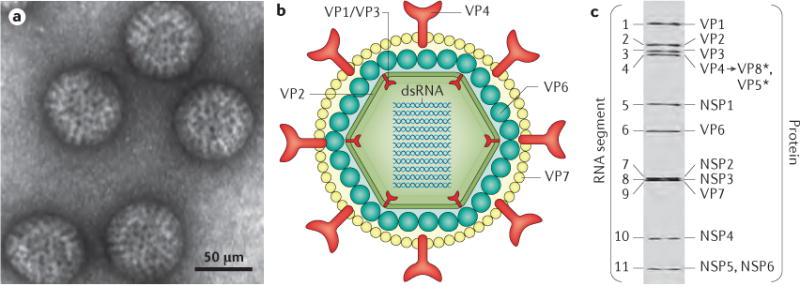
    1. Bishop RF, et al. Virus particles in epithelial cells of duodenal mucosa from children with acute non-bacterial gastroenteritis. Lancet. 1973;2:1281–1283. This study shows visualization of rotavirus as the causative agent of viral gastroenteritis and the associated histopathology. - PubMed
    1. Flewett TH, et al. Letter: Virus particles in gastroenteritis. Lancet. 1973;2:1497. - PubMed
    1. Estes MK, Greenberg HB. In: Field’s Virology. Knipe DM, Howley PM, editors. Lippincott: Williams & Wilkins; 2013. pp. 1347–1401.
    1. Matthijnssens J, et al. VP6-sequence-based cutoff values as a criterion for rotavirus species demarcation. Arch Virol. 2012;157:1177–1182. - PubMed

MeSH terms