. 2017 Nov 9;8(11):e3167.
doi: 10.1038/cddis.2017.520.

Bortezomib-induced miRNAs direct epigenetic silencing of locus genes and trigger apoptosis in leukemia

Affiliations

Bortezomib-induced miRNAs direct epigenetic silencing of locus genes and trigger apoptosis in leukemia

Yu-Yi Chu et al. Cell Death Dis. .

Abstract

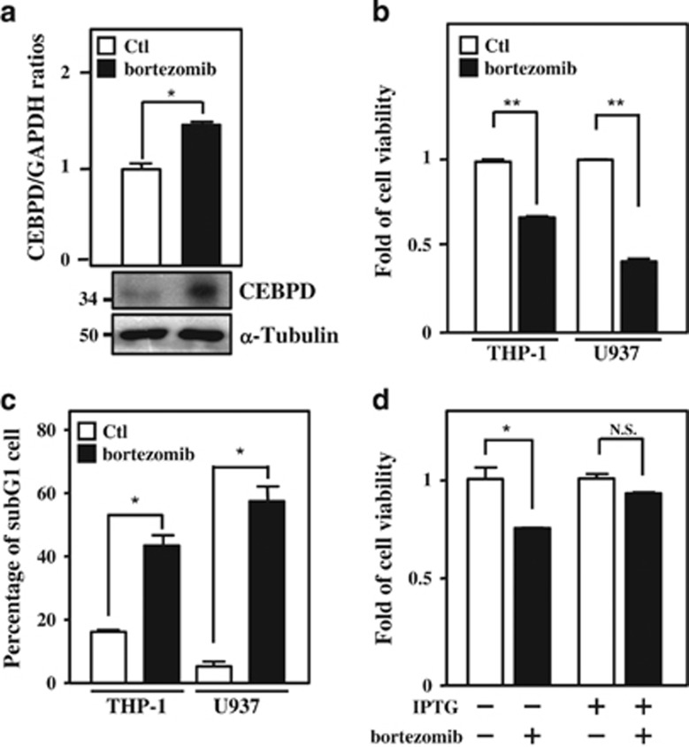
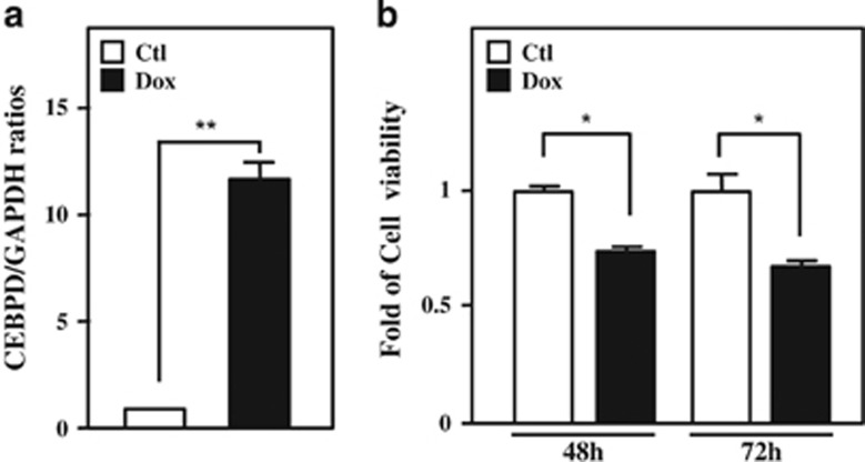
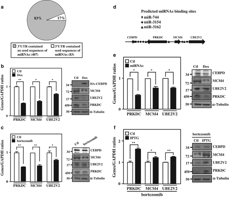
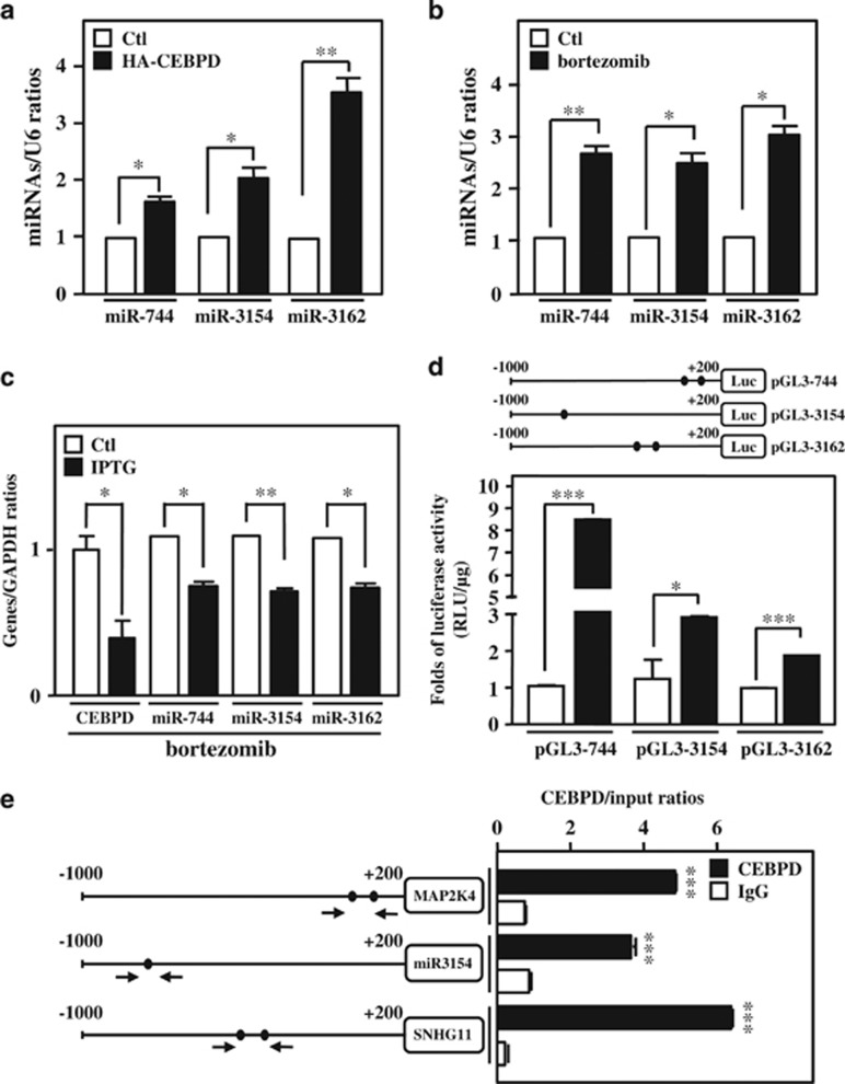
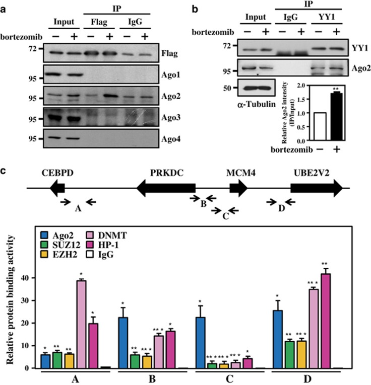
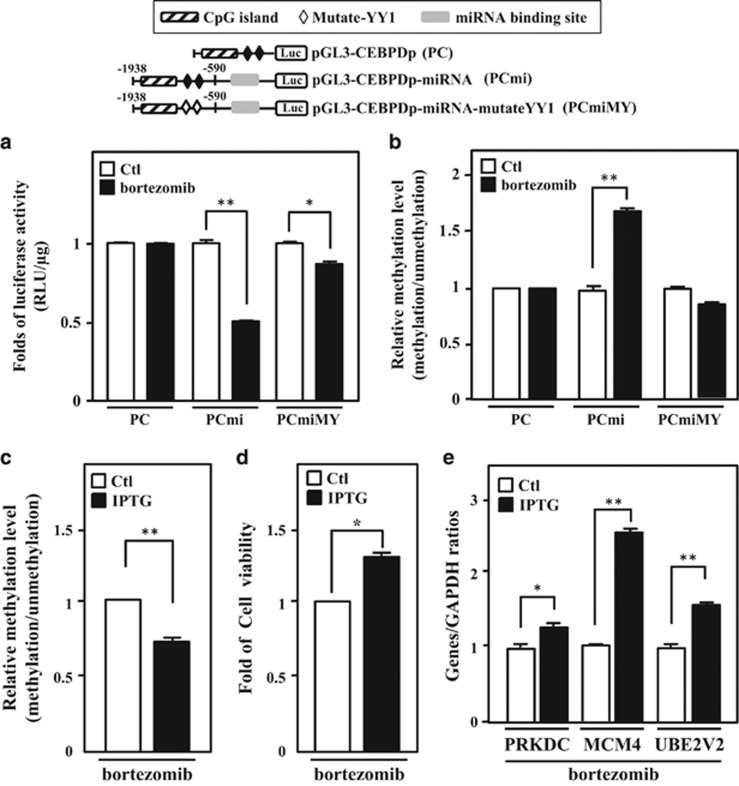
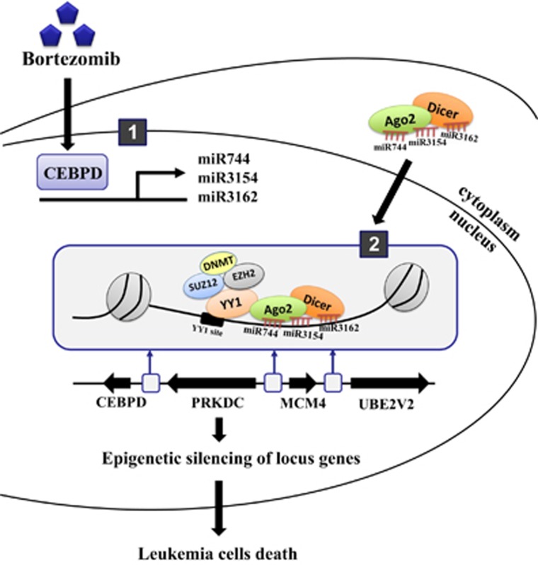
MicroRNAs (miRNAs) have been suggested to repress transcription via binding the 3'-untranslated regions of mRNAs. However, the involvement and details of miRNA-mediated epigenetic regulation, particularly in targeting genomic DNA and mediating epigenetic regulation, remain largely uninvestigated. In the present study, transcription factor CCAAT/enhancer binding protein delta (CEBPD) was responsive to the anticancer drug bortezomib, a clinical and highly selective drug for leukemia treatment, and contributed to bortezomib-induced cell death. Interestingly, following the identification of CEBPD-induced miRNAs, we found that miR-744, miR-3154 and miR-3162 could target CpG islands in the 5'-flanking region of the CEBPD gene. We previously demonstrated that the Yin Yang 1 (YY1)/polycomb group (PcG) protein/DNA methyltransferase (DNMT) complex is important for CCAAT/enhancer binding protein delta (CEBPD) gene inactivation; we further found that Argonaute 2 (Ago2) interacts with YY1 and binds to the CEBPD promoter. The miRNA/Ago2/YY1/PcG group protein/DNMT complex linked the inactivation of CEBPD and genes adjacent to its 5'-flanking region, including protein kinase DNA-activated catalytic polypeptide (PRKDC), minichromosome maintenance-deficient 4 (MCM4) and ubiquitin-conjugating enzyme E2 variant 2 (UBE2V2), upon bortezomib treatment. Moreover, we revealed that miRNA binding is necessary for YY1/PcG group protein/DNMT complex-mediated epigenetic gene silencing and is associated with bortezomib-induced methylation on genomic DNA. The present study successfully characterized the interactions of the miRNA/Ago2/YY1/PcG group protein/DNMT complex and provided new insights for miRNA-mediated epigenetic regulation in bortezomib-induced leukemic cell arrest and cell death.


Conflict of interest statement

The authors declare no conflict of interest.

Figures

Figure 1
Figure 1
CEBPD, which contributes to leukemic cell apoptosis, is responsive to bortezomib. (a) Bortezomib increases the expression of CEBPD. After treatment with bortezomib (50 nM) for 6 h in THP-1 cells, total RNA and protein lysates were harvested and analyzed using qPCR and western blot analysis. (b) Bortezomib inhibits cell viability. After treatment with bortezomib (50 nM) for 24 h, the viability of leukemic cells (THP-1 and U937) was measured using an MTT assay. (c) Bortezomib induces the growth arrest of leukemic cells. After treating with bortezomib (50 nM) for 24 h, the sub-G1 phase of THP-1 and U937 cells was analyzed by flow cytometry. (d) Attenuated CEBPD reverses bortezomib-reduced cell viability in THP-1 cells. THP-1 cells with the IPTG-inducible CEBPD knockdown system were pretreated with IPTG (500 μM) for 3 h. After treatment with bortezomib (50 nM) for 24 h, cell viability was measured using an MTT assay. The data are presented as the mean±standard error of experiments performed in triplicate (*P<0.05, **P<0.01, Student’s t-test). NS, not significant
Figure 2
Figure 2
CEBPD inhibits the survival rates of THP-1 cells. (a) Dox successfully induces CEBPD expression in THP-1 cells in the Dox-inducible CEBPD expression system. THP-1 cells were treated with Dox (1 μg/ml) for 6 h, followed by qPCR to analyze the expression of CEBPD. (b) CEBPD reduces the viability of THP-1 cells. THP-1 cells with the Dox-inducible CEBPD expression system were treated with Dox (1 μg/ml), and cell viability was measured using an MTT assay. The data are presented as the mean±standard error of experiments performed in triplicate (*P<0.05, **P<0.01, Student’s t-test). NS, not significant
Figure 3
Figure 3
CEBPD and responsive miRNAs contribute to the inactivation of the surrounding genes. (a) The 3′-UTRs of most CEBPD-downregulated mRNAs are not targeted by CEBPD-upregulated miRNAs. The CEBPD-upregulated miRNAs and -downregulated mRNAs microarray profiling was analyzed using the miRTar prediction program. The pie chart presents the percentage of CEBPD-downregulated mRNAs in the 3′-UTR, containing the seed sequences of CEBPD-upregulated miRNAs. (b) CEBPD participates in the inactivation of the surrounding genes. THP-1 cells with the Dox-inducible CEBPD expression system were treated with Dox (1 μg/ml) for 18 h; total RNA was harvested to perform qPCR (left panel), and total protein was harvested to perform western blot analysis (right panel). (c) Bortezomib represses the expression of PRKDC, MCM4 and UBE2V2. After treating with bortezomib (50 nM) for 16 h, total RNA was harvested to perform qPCR (left panel) and total protein was harvested to perform the western blot analysis (right panel). (d) Schematic representation showing the location of genes and putative miRNA-binding motifs. (e) miR-744, miR-3154 and miR-3162 reduces CEBPD surrounding genes. THP-1 cells with the Dox-inducible miR-744, miR-3154 and miR-3162 expression system were treated with Dox (1 μg/ml) for 18 h. Total RNA was harvested to perform qPCR (left panel), and total protein was harvested to perform the western blot analysis (right panel). (f) Attenuated CEBPD reverses bortezomib-inhibited gene expression in THP-1 cells. THP-1 cells with the IPTG-inducible CEBPD knockdown system were pretreated with IPTG (500 μM) for 3 h. After treatment with bortezomib (50 nM) for 16 h, total RNA was harvested to perform qPCR (left panel) and total protein was harvested to perform the western blot analysis (right panel). The data are presented as the mean±standard error of experiments performed in triplicate (*P<0.05, **P<0.01, Student’s t-test)
Figure 4
Figure 4
miR-744, miR-3154 and miR-3162 are CEBPD-responsive miRNAs. (a) CEBPD induces miR-744, miR-3154 and miR-3162 expression. THP-1 cells were transfected with HA-CEBPD for 18 h. Total RNA was harvested, and the levels of miRNAs (miR-744, miR-3154 and miR-3162) were confirmed by qPCR. (b) Bortezomib increases miR-744, miR-3154 and miR-3162 expression. qPCR was performed using total RNA harvested from THP-1 cells treated with bortezomib (50 nM) for 6 h. (c) The loss of CEBPD reduced bortezomib-induced miR-744, miR-3154 and miR-3162. THP-1 cells with IPTG-inducible CEBPD knockdown system were pretreated with IPTG (500 μM) for 3 h. After treatment with bortezomib (50 nM) for 6 h, the expression levels of miR-744, miR-3154 and miR-3162 were measured using qPCR. (d) CEBPD activates the promoter activity of miRNAs. Schematic representation of the reporter constructs with the miR-744, miR-3154 and miR-3162 promoters. The approximate location of putative CEBPD-binding motifs is indicated with an oval. The luciferase activity was assessed after co-transfecting reporters and expression vectors in THP-1 cells as indicated. (e) CEBPD directly binds to the miR-744, miR-3154 and miR-3162 promoters in vivo. A ChIP assay was performed in THP-1 cells with the Dox-inducible CEBPD expression system. Sonicated chromatin was subjected to ChIP-qPCR analysis using CEBPD or control IgG antibodies. The data are presented as the mean±standard error of experiments performed in triplicate (*P<0.05, **P<0.01, ***P<0.001, Student’s t-test)
Figure 5
Figure 5
Bortezomib induces the formation of the Ago2/YY1 complex and the binding of the PcG complex. (a and b) Bortezomib induces Ago2 and YY1 interaction. The 293T cells were transfected with Flag-YY1 and treated with bortezomib (50 nM) for 5 h. Total lysates were collected and analyzed using immunoprecipitation using an anti-Flag antibody (a). The THP-1 cells were treated with bortezomib (50 nM) for 12 h. Total lysates were collected and analyzed using immunoprecipitation using an anti-YY1 antibody (b, upper panel), and quantitated results showed the interaction intensity between YY1 and Ago2 (b, bottom panel). (c) The Ago2-PcG complex (SUZ12, DNMT, EZH2) and HP-1 directly bind to the upstream region of CEBPD and the surrounding genes. A ChIP assay was performed using THP-1 cells treated with bortezomib (50 nM). The sonicated chromatin of THP-1 cells was separately immunoprecipitated using specific antibodies against Ago2, SUZ12, DNMT, EZH2, HP-1 and control IgG. The immunoprecipitated DNA was qPCR amplified using different primers as indicated in the top panel. The data are presented as the mean±standard error of experiments performed in triplicate (*P<0.05, **P<0.01, ***P<0.001, Student’s t-test)
Figure 6
Figure 6
miR-744, miR-3154 and miR-3162 mediates a TGS through a direct binding on gDNA. (a) The binding of miRNAs and YY1 is associated with bortezomib-induced epigenetic gene silencing and methylation level. miR-744, miR-3154 and miR-3162 are necessary for YY1/PcG complex-mediated epigenetic gene silencing. Schematic representation of reporter constructs with the CEBPD promoter only (PC), CEBPD promoter constructed with miRNAs binding sites (PCmi) and PCmi with mutated YY1 binding sites (PCmiMY). The luciferase activity was assessed after transfecting various constructs, as indicated, in 293T cells and subsequently treating the cells with bortezomib (50 nM). (b) The binding of miRNAs and YY1 increase the methylation levels of the CpG islands on CEBPD promoters. After transfecting constructs into 293T with or without bortezomib (50 nM) treatment, the methylation level of CpG islands on the reporters (PC, PCmi and PCmiMY) was obtained using quantitative methylation-specific PCR. (c) miR-744, miR-3154 and miR-3162 are critical for bortezomib-induced DNA methylation. After transfecting constructs into 293T with the IPTG-inducible miR-744/3154/3162-silencing system, the cells were pretreated with IPTG (500 μM) for 3 h and treated with bortezomib (50 nM) for 16 h. Methylation level of CpG islands on the reporter (PCmi) was obtained using quantitative methylation-specific PCR. (d) Inhibition of miR-744, miR-3154 and miR-3162 reverses bortezomib-reduced cell viability in THP-1 cells. THP-1 cells with the IPTG-inducible miRNA-744/3154/3162-silencing system were pretreated with IPTG (500 μM) for 3 h. After treating with bortezomib (50 nM) for 24 h, cell viability was measured by the MTT assay. (e) Attenuated miR-744, miR-3154 and miR-3162 reverses bortezomib-reduced PRKDC, MCM4 and UBE2V2 expression. THP-1 cells with the IPTG-inducible miR-744/3154/3162-silencing system were pretreated with IPTG (500 μM) for 3 h. After treating with bortezomib (50 nM) for 16 h, the expression levels of PRKDC, MCM4 and UBE2V2 were measured by qPCR. The data are presented as the mean±standard error of experiments performed in triplicate (*P<0.05, **P<0.01, Student’s t-test)
Figure 7
Figure 7
Schematic model for CEBPD turned off the locus genes and resulted in leukemic cell death through miRNA/Ago2/YY1/PcG group protein/DNMT complex-directed epigenetic silencing. In response to the anticancer drug bortezomib, the transcription factor CEBPD is activated and transcriptionally activates miR-744, miR-3154 and miR-3162. These miRNAs form a complex with Ago2 and translocate into the nucleus to target their complementary DNA sequence-binding sites on the promoter regions of CEBPD, PRKDC, MCM4 and UBE2V2. The initiator miRNAs/Ago2 complex interacts with YY1 and recruits the epigenetic regulators, the PcG complex/DNMTs, to silence the four gene loci, including CEBPD itself. The inactivation of these potent oncogenes, PRKDC, MCM4 and UBE2V2 result in leukemic cell death via CEBPD-responsive miRNA-mediated epigenetic silencing

Similar articles

Cited by

References

    1. He L, Hannon GJ. MicroRNAs: small RNAs with a big role in gene regulation. Nat Rev Genet 2004; 5: 522–531. - PubMed
    1. Garzon R, Marcucci G, Croce CM. Targeting microRNAs in cancer: rationale, strategies and challenges. Nat Rev Drug Discov 2010; 9: 775–789. - PMC - PubMed
    1. Hammond SM, Bernstein E, Beach D, Hannon GJ. An RNA-directed nuclease mediates post-transcriptional gene silencing in Drosophila cells. Nature 2000; 404: 293–296. - PubMed
    1. Ghildiyal M, Zamore PD. Small silencing RNAs: an expanding universe. Nat Rev Genet 2009; 10: 94–108. - PMC - PubMed
    1. Younger ST, Pertsemlidis A, Corey DR. Predicting potential miRNA target sites within gene promoters. Bioorg Med Chem Lett 2009; 19: 3791–3794. - PMC - PubMed

MeSH terms