

In vitro characterization of gamma oscillations in the hippocampal formation of the domestic chick
- PMID: 29120510
- PMCID: PMC6220815
- DOI: 10.1111/ejn.13773
In vitro characterization of gamma oscillations in the hippocampal formation of the domestic chick
Abstract
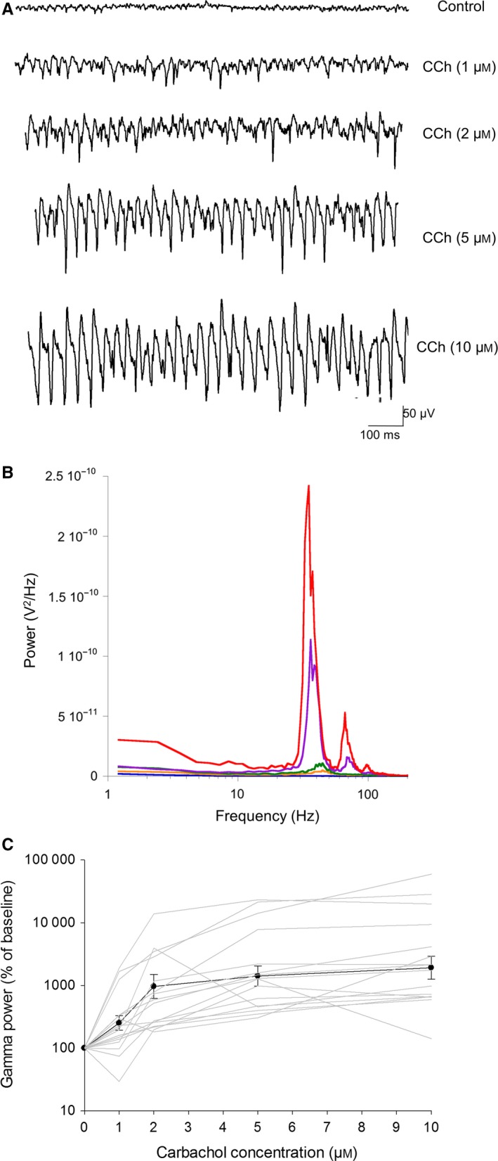
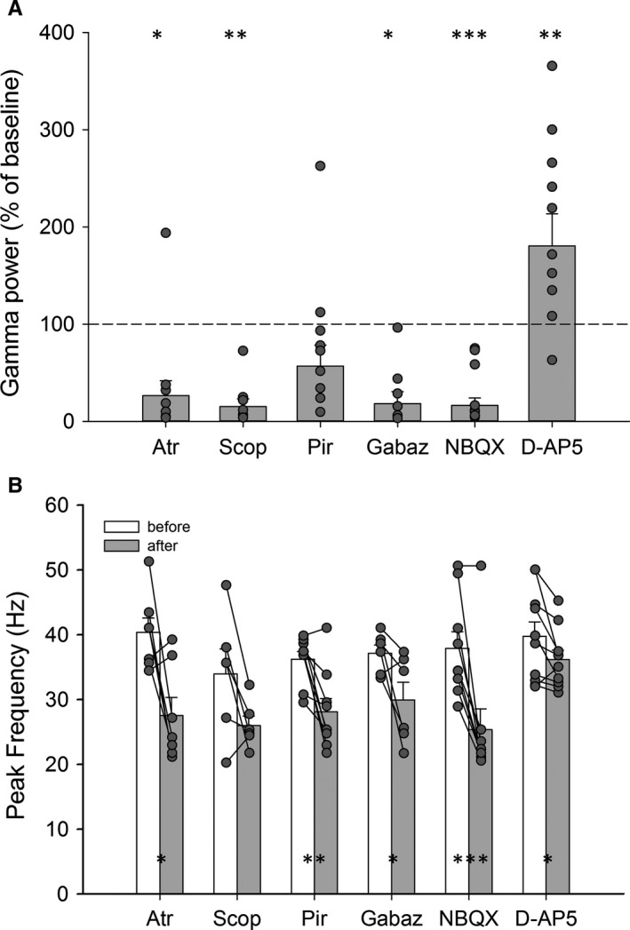
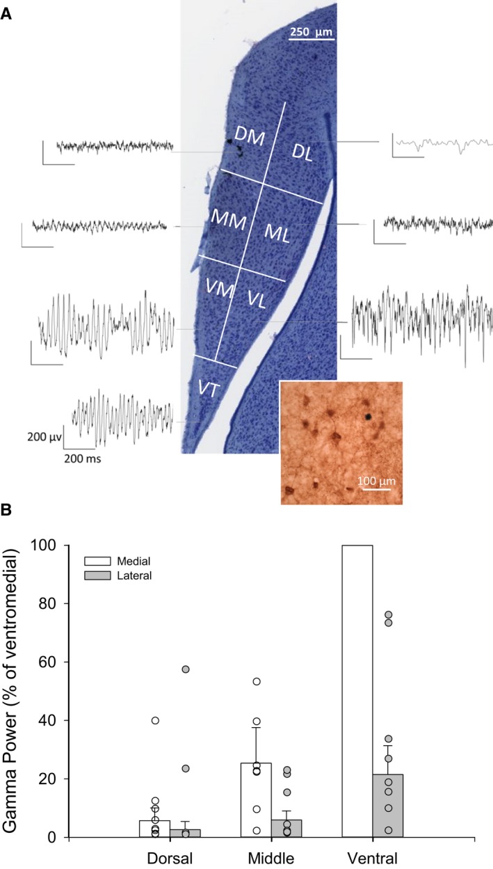
Avian and mammalian brains have evolved independently from each other for about 300 million years. During that time, the hippocampal formation (HF) has diverged in morphology and cytoarchitecture, but seems to have conserved much of its function. It is therefore an open question how seemingly different neural organizations can generate the same function. A prominent feature of the mammalian hippocampus is that it generates different neural oscillations, including the gamma rhythm, which plays an important role in memory processing. In this study, we investigate whether the avian hippocampus also generates gamma oscillations, and whether similar pharmacological mechanisms are involved in this function. We investigated the existence of gamma oscillations in avian HF using in vitro electrophysiology in P0-P12 domestic chick (Gallus gallus domesticus) HF brain slices. Persistent gamma frequency oscillations were induced by the bath application of the cholinergic agonist carbachol, but not by kainate, a glutamate receptor agonist. Similar to other species, carbachol-evoked gamma oscillations were sensitive to GABAA , AMPA/kainate and muscarinic (M1) receptor antagonism. Therefore, similar to mammalian species, muscarinic receptor-activated avian HF gamma oscillations may arise via a pyramidal-interneuron gamma (PING)-based mechanism. Gamma oscillations are most prominent in the ventromedial area of the hippocampal slices, and gamma power is reduced more laterally and dorsally in the HF. We conclude that similar micro-circuitry may exist in the avian and mammalian hippocampal formation, and this is likely to relate to the shared function of the two structures.
Keywords: Gallus gallus domesticus; avian hippocampus; homology; local field potentials; rhythmogenesis.
© 2017 The Authors. European Journal of Neuroscience published by Federation of European Neuroscience Societies and John Wiley & Sons Ltd.
Figures
References
-
- Andersen, P. (2007). The Hippocampus Book. Oxford University Press, Cary, NC.
-
- Atoji, Y. & Wild, J.M. (2004) Fiber connections of the hippocampal formation and septum and subdivisions of the hippocampal formation in the pigeon as revealed by tract tracing and kainic acid lesions. J. Comp. Neurol., 475, 426–461. - PubMed
-
- Atoji, Y. & Wild, J.M. (2006) Anatomy of the avian hippocampal formation. Rev. Neuroscience, 17, 3–15. - PubMed
Publication types
MeSH terms
Substances
Grants and funding
LinkOut - more resources
Full Text Sources
Other Literature Sources
Molecular Biology Databases
Research Materials
Miscellaneous
