. 2017 Nov 8;22(5):653-666.e5.
doi: 10.1016/j.chom.2017.10.006.

Staphylococcus aureus Epicutaneous Exposure Drives Skin Inflammation via IL-36-Mediated T Cell Responses

Affiliations

Staphylococcus aureus Epicutaneous Exposure Drives Skin Inflammation via IL-36-Mediated T Cell Responses

Haiyun Liu et al. Cell Host Microbe. .

Abstract

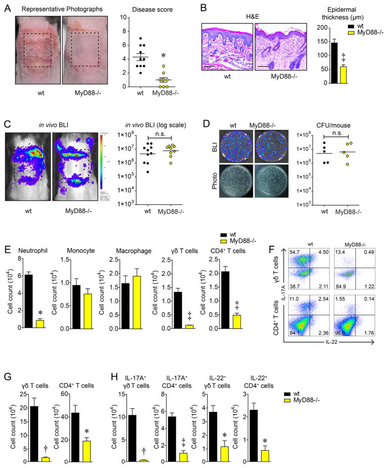
Staphylococcus aureus colonization contributes to skin inflammation in diseases such as atopic dermatitis, but the signaling pathways involved are unclear. Herein, epicutaneous S. aureus exposure to mouse skin promoted MyD88-dependent skin inflammation initiated by IL-36, but not IL-1α/β, IL-18, or IL-33. By contrast, an intradermal S. aureus challenge promoted MyD88-dependent host defense initiated by IL-1β rather than IL-36, suggesting that different IL-1 cytokines trigger MyD88 signaling depending on the anatomical depth of S. aureus cutaneous exposure. The bacterial virulence factor PSMα, but not α-toxin or δ-toxin, contributed to the skin inflammation, which was driven by IL-17-producing γδ and CD4+ T cells via direct IL-36R signaling in the T cells. Finally, adoptive transfer of IL-36R-expressing T cells to IL-36R-deficient mice was sufficient for mediating S. aureus-induced skin inflammation. Together, this study defines a previously unknown pathway by which S. aureus epicutaneous exposure promotes skin inflammation involving IL-36R/MyD88-dependent IL-17 T cell responses.

Keywords: IL-17; IL-36; Staphylococcus aureus; T cell; atopic dermatitis; cutaneous; epicutaneous; inflammation; phenol soluble modulin; skin.


Figures

Figure 1
Figure 1
MyD88−/− mice develop decreased skin inflammation after epicutaneous S. aureus exposure. Wt and MyD88−/− mice were epicutaneously challenged with S. aureus (1×10⁸ CFU) on the dorsal skin for 7 days. (A) Representative skin photographs and mean disease score ± s.e.m. (B) Representative histology (hematoxylin and eosin [H&E] stain, 200× magnification) and mean epidermal thickness ± s.e.m. (C) Representative in vivo bioluminescent imaging (BLI) signals and total flux (photons/s) (log10 scale). (D) Representative bacterial culture plates after overnight culture ± BLI and CFU (log10 scale). (E) Mean cell number ± s.e.m. from the skin as measured by FACS. (F) Representative flow plots and (G,H) mean cell number ± s.e.m. of total and IL-17A- and IL-22-producing T cells from dLNs as measured by FACS. *P<0.05, †P<0.01, ‡ P<0.001 (MyD88−/− mice versus wt mice) as calculated by two-tailed Student’s t-test. n.s. = not significant. Results are representative of at least 2 independent experiments.
Figure 2
Figure 2
IL-36R/MyD88 signaling by T cells mediates S. aureus-induced skin inflammation. Mice were epicutaneously challenged with S. aureus (1×10⁸ CFU) on the dorsal skin for 7 days. (A) Representative skin photographs for wt versus K14CreER×MyD88fl/fl (K14-MyD88−/−), LysMCre×MyD88fl/fl (LysM-MyD88−/−), LckCre×MyD88fl/fl (Lck-MyD88−/−) mice, and MyD88−/− mice. (B) Mean disease score ± s.e.m. (C) Mean epidermal thickness ± s.e.m. (D,E) Wt C57BL/6 versus IL-1α−/−, IL-β−/−, IL-18R1−/−, and IL-36R−/− mice mean disease score ± s.e.m. and mean epidermal thickness ± s.e.m. (F,G) Wt Balb/c versus IL-33−/− mice mean disease score ± s.e.m. and mean epidermal thickness ± s.e.m. *P<0.05, †P<0.01, ‡ P<0.001 as calculated by two-tailed Student’s t-test. Results are representative of at least 2 independent experiments. See also Figure S1 and S2.
Figure 3
Figure 3
PSMα plays major role in mediating skin inflammation during S. aureus epicutaneous exposure. Wt C57/BL6 mice were epicutaneously challenged with PBS, S. aureus USA300 LAC parent, δ-toxin, α-toxin, PSMα mutant (Δ) (1×10⁸ CFU) strains, and S. epidermidis (1×10⁸ CFU) on dorsal skin for 7 days. (A,D,G) Representative skin photographs. (B,E,H) Mean disease score ± s.e.m. (C,F,I). Representative histology (hematoxylin and eosin [H&E] stain, 200× magnification) and mean epidermal thickness ± s.e.m. *P<0.05, ‡ P<0.001 as calculated by two-tailed Student’s t-test. Results are representative of at least 2 independent experiments.
Figure 4
Figure 4
The trigger for MyD88-signaling is different dependent upon the depth of exposure to S. aureus in the skin. Mice were infected with S. aureus (3×10⁷ CFU) through i.d. injection on dorsal skin, and the skin lesion and bacterial burden were monitored for 14 days in wt versus IL-β−/− and IL-36R−/− mice. (A,B) Representative skin photographs and quantitative total lesion area (cm2) ± s.e.m. (C,D) Representative in vivo bioluminescent imaging (BLI) signals and mean total flux (photons/s) ± s.e.m. (log10 scale). ‡ P<0.001 (IL-β−/− mice versus wt mice) as calculated by two-way ANOVA. (E,F) Mean levels of mRNA in arbitrary units (A.U.) ± s.e.m. of IL-1β and IL-36α from skin tissue homogenates at 0, 3, 7 days after S. aureus epicutaneous exposure (1×10⁸ CFU) or intradermal infection (3×10⁷ CFU). *P<0.05, ‡ P<0.001 as calculated by two-tailed Student’s t-test. Results are representative of at least 2 independent experiments.
Figure 5
Figure 5
IL-36R−/− mice have impaired IL-17A and IL-22 T cell responses. Wt and IL-36R−/− mice were epicutaneously challenged with S. aureus (1×10⁸ CFU) on the dorsal skin for 7 days. (A) mRNA levels of IL-36α, IL-36β, and IL-36γ relative to β-actin, on day 3 and 7 after S. aureus challenge, measured by RT-PCR ± s.e.m. (B) Representative immunofluorescence of skin sections labeled with anti-IL-36α (green, upper panels) and merged with DAPI (blue, lower panels), 400× magnification. Dashed line = dermoepidermal junction. (C–E) Protein array analysis of IL-17A, IL-22, IFN-γ, IL-13, and IL-33 protein concentration ± s.e.m. in wt and IL-36R−/− mice lesional skin. (F–I) Representative flow plots and mean cell number ± s.e.m. of total and IL-17A-, IL-22-producing T cells from dLNs as measured by FACS. *P<0.05, †P<0.01, ‡ P<0.001 (IL-36R−/− mice versus wt mice) as calculated by two-tailed Student’s t-test. n.s. = not significant. Results are representative of at least 2 independent experiments.
Figure 6
Figure 6
IL-17A/F−/− mice have decreased skin inflammation after epicutaneous S. aureus exposure. Mice were epicutaneously challenged with S. aureus (1×10⁸ CFU) on the dorsal skin for 7 days in wt versus IL-17A/F−/− and IL-22−/− mice. (A) Mean disease score ± s.e.m. (B) Mean epidermal thickness ± s.e.m. (C,D) Mean cell number ± s.e.m. of IL-17A-producing cells including γδ T cells, CD4+ T cells, CD8+ T cells, ILC3s (CD45+ CD127+ RORγt+, Lin), B cells (B220+), NK cells (NK1.1+), and myeloid cells (CD11b+) from skin and dLNs as measured by FACS. *P<0.05 (IL-17A/F−/− mice versus wt mice) as calculated by two-tailed Student’s t-test. Results are representative of at least 2 independent experiments.
Figure 7
Figure 7
IL-36R-signaling by T cells directly promotes IL-17A production and skin inflammation. Naïve CD3+ T cells purified from wt or IL-36R−/− mice skin dLNs were co-cultured in vitro for 72h with CD11c+ DCs from naïve wt or IL-36R−/− mice pulsed with heat-killed S. aureus ± exogenous IL-36α. (A,B) Representative flow plots and mean cell number ± s.e.m. of total and IL-17A-, IL-22-producing T cells in the in vitro culture as measured by FACS. (C) 3×10⁶ naïve CD3+ T cells purified from wt or IL-36R−/− mice skin dLNs were adoptively transferred into wt or IL-36R−/− mice; after 7 days of rest, mice were colonized with S. aureus (1×10⁸ CFU) for 7 days. Mean disease score ± s.e.m., mean epidermal thickness ± s.e.m. and lesional skin IL-17A protein concentration ± s.e.m. *P<0.05, †P<0.01, ‡ P<0.001 as calculated by two-tailed Student’s t-test. n.s. = not significant. Results are representative of at least 2 independent experiments. See also Figure S3 and S4.

Comment in

References

    1. Breuer K, SHA, Kapp A, Werfel T. Staphylococcus aureus: colonizing features and influence of an antibacterial treatment in adults with atopic dermatitis. The British journal of dermatology. 2002;147:55–61. - PubMed
    1. Brown AF, Leech JM, Rogers TR, McLoughlin RM. Staphylococcus aureus Colonization: Modulation of Host Immune Response and Impact on Human Vaccine Design. Frontiers in immunology. 2014;4:507. - PMC - PubMed
    1. Byrd AL, Deming C, Cassidy SKB, Harrison OJ, Ng WI, Conlan S, Program NCS, Belkaid Y, Segre JA, Kong HH. Staphylococcus aureus and Staphylococcus epidermidis strain diversity underlying pediatric atopic dermatitis. Science translational medicine. 2017:9. - PMC - PubMed
    1. Cai Y, Shen X, Ding C, Qi C, Li K, Li X, Jala VR, Zhang HG, Wang T, Zheng J, et al. Pivotal role of dermal IL-17-producing gammadelta T cells in skin inflammation. Immunity. 2011;35:596–610. - PMC - PubMed
    1. Carrier Y, Ma HL, Ramon HE, Napierata L, Small C, O’Toole M, Young DA, Fouser LA, Nickerson-Nutter C, Collins M, et al. Inter-regulation of Th17 cytokines and the IL-36 cytokines in vitro and in vivo: implications in psoriasis pathogenesis. The Journal of investigative dermatology. 2011;131:2428–2437. - PubMed

MeSH terms