

Indocyanine green fluorescence in second near-infrared (NIR-II) window
- PMID: 29121078
- PMCID: PMC5679521
- DOI: 10.1371/journal.pone.0187563
Indocyanine green fluorescence in second near-infrared (NIR-II) window
Abstract
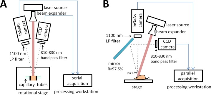
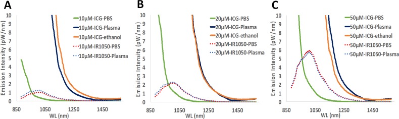
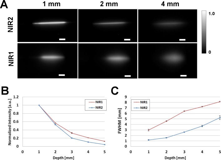
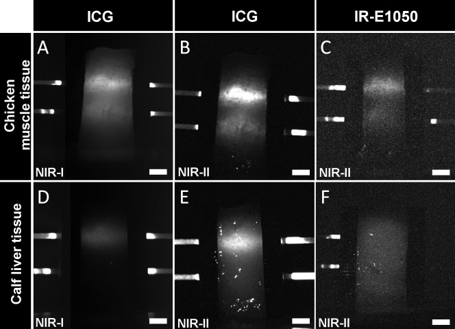
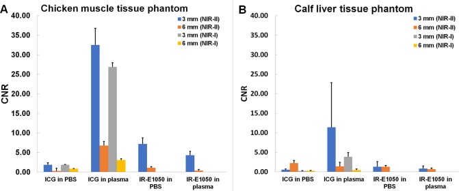
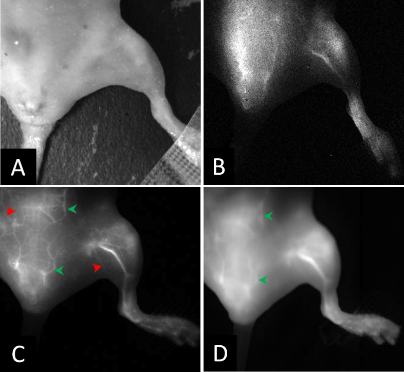
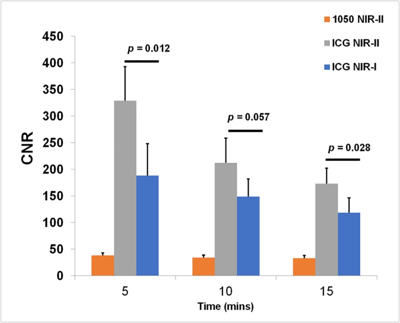
Indocyanine green (ICG), a FDA approved near infrared (NIR) fluorescent agent, is used in the clinic for a variety of applications including lymphangiography, intra-operative lymph node identification, tumor imaging, superficial vascular imaging, and marking ischemic tissues. These applications operate in the so-called "NIR-I" window (700-900 nm). Recently, imaging in the "NIR-II" window (1000-1700 nm) has attracted attention since, at longer wavelengths, photon absorption, and scattering effects by tissue components are reduced, making it possible to image deeper into the underlying tissue. Agents for NIR-II imaging are, however, still in pre-clinical development. In this study, we investigated ICG as a NIR-II dye. The absorbance and NIR-II fluorescence emission of ICG were measured in different media (PBS, plasma and ethanol) for a range of ICG concentrations. In vitro and in vivo testing were performed using a custom-built spectral NIR assembly to facilitate simultaneous imaging in NIR-I and NIR-II window. In vitro studies using ICG were performed using capillary tubes (as a simulation of blood vessels) embedded in Intralipid solution and tissue phantoms to evaluate depth of tissue penetration in NIR-I and NIR-II window. In vivo imaging using ICG was performed in nude mice to evaluate vascular visualization in the hind limb in the NIR-I and II windows. Contrast-to-noise ratios (CNR) were calculated for comparison of image quality in NIR-I and NIR-II window. ICG exhibited significant fluorescence emission in the NIR-II window and this emission (similar to the absorption profile) is substantially affected by the environment of the ICG molecules. In vivo imaging further confirmed the utility of ICG as a fluorescent dye in the NIR-II domain, with the CNR values being ~2 times those in the NIR-I window. The availability of an FDA approved imaging agent could accelerate the clinical translation of NIR-II imaging technology.
Conflict of interest statement
Figures
References
-
- Landsman ML, Kwant G, Mook GA, Zijlstra WG. Light-absorbing properties, stability, and spectral stabilization of indocyanine green. J Appl Physiol. 1976;40: 575–583. - PubMed
-
- FOX IJ, WOOD EH. Indocyanine green: physical and physiologic properties. Proc Staff Meet Mayo Clin. 1960;35: 732–744. - PubMed
-
- Flower RW, Hochheimer BF. Indocyanine green dye fluorescence and infrared absorption choroidal angiography performed simultaneously with fluorescein angiography. Johns Hopkins Med J. 1976;138: 33–42. - PubMed
-
- Reuthebuch O, Haussler A, Genoni M, Tavakoli R, Odavic D, Kadner A, et al. Novadaq SPY*. CHEST. The American College of Chest Physicians; 2004;125: 418–424. doi: 10.1378/chest.125.2.418 - DOI - PubMed
-
- Hirata A, Tanihara H. Ruptured internal limiting membrane associated with blunt trauma revealed by indocyanine green staining. Graefe's Arch Clin Exp Ophthalmol. 2004;242: 527–530. doi: 10.1007/s00417-004-0875-1 - DOI - PubMed
MeSH terms
Substances
LinkOut - more resources
Full Text Sources
Other Literature Sources
Miscellaneous
