

The Mouse Model of Pancreatic Cancer Atlas (MMPCA) for classification of pancreatic cancer lesions: A large histological investigation of the Ptf1aCre/+;LSL-KrasG12D/+ transgenic mouse model of pancreatic cancer
- PMID: 29121082
- PMCID: PMC5679567
- DOI: 10.1371/journal.pone.0187552
The Mouse Model of Pancreatic Cancer Atlas (MMPCA) for classification of pancreatic cancer lesions: A large histological investigation of the Ptf1aCre/+;LSL-KrasG12D/+ transgenic mouse model of pancreatic cancer
Abstract
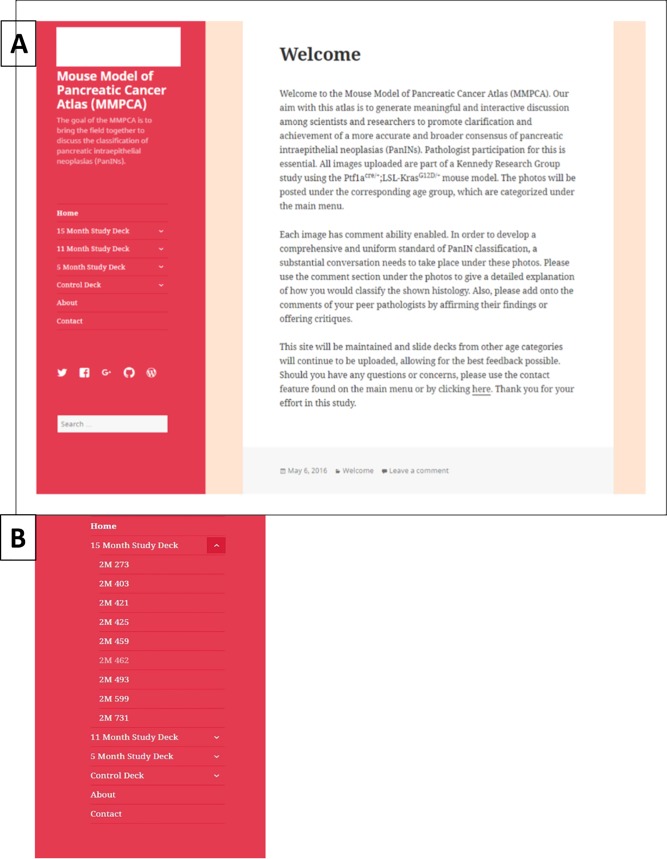
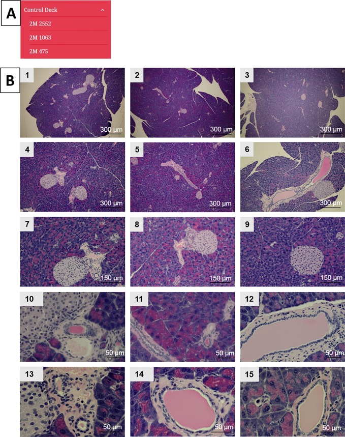
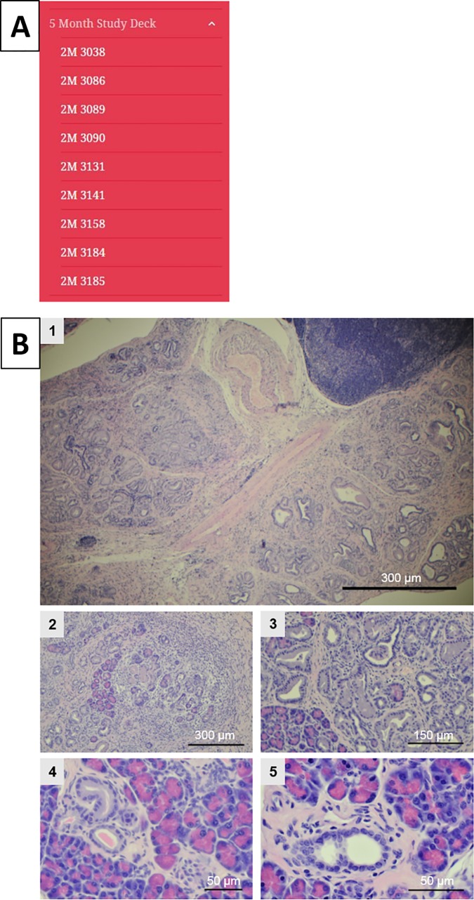
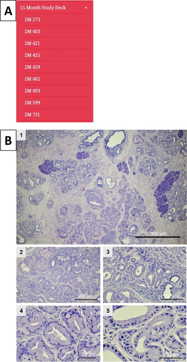
Pancreatic ductal adenocarcinoma (PDAC) is one of the leading forms of cancer related deaths in the United States. With limited treatment options and unreliable diagnostic methods, long-term survival rates following a diagnosis of pancreatic cancer remain poor. Pancreatic intraepithelial neoplasia (PanIN) are precancerous lesions that precede progression towards PDAC. PanIN occur in increasing complexity as the disease progresses and the description of PanIN plays a critical role in describing, staging and diagnosing PDAC. Inconsistencies in PanIN classifications exist even amongst leading pathologists. This has led to debate and confusion among researchers and pathologists involved in pancreatic cancer research, diagnosis and treatment. We have sought to initiate a discussion with leading pathologists with a goal of increasing consensus in the interpretation of PanIN and associated structures within the precancerous pancreas. Toward achieving this goal, we are in the process of conducting an extensive study of over 1000 male and female pancreata in varying stages of PanIN progression isolated from the Ptf1aCre/+;LSL-KrasG12D/+ transgenic mouse model of pancreatic cancer. Using this extensive database, we have established the Mouse Model of Pancreatic Cancer Atlas (MMPCA) to serve as a platform for meaningful and interactive discussion among researchers and pathologists who study pancreatic disease. We hope that the MMPCA will be an effective tool for promoting a more consistent and accurate consensus of PanIN classifications in the future.
Conflict of interest statement
Figures

Similar articles
-
Calorie restriction delays the progression of lesions to pancreatic cancer in the LSL-KrasG12D; Pdx-1/Cre mouse model of pancreatic cancer.Exp Biol Med (Maywood). 2013 Jul;238(7):787-97. doi: 10.1177/1535370213493727. Epub 2013 Jul 4. Exp Biol Med (Maywood). 2013. PMID: 23828595
-
HMGA1 expression levels are elevated in pancreatic intraepithelial neoplasia cells in the Ptf1a-Cre; LSL-KrasG12D transgenic mouse model of pancreatic cancer.Br J Cancer. 2017 Aug 22;117(5):639-647. doi: 10.1038/bjc.2017.216. Epub 2017 Jul 11. Br J Cancer. 2017. PMID: 28697176 Free PMC article.
-
Requirement of NEMO/IKKγ for effective expansion of KRAS-induced precancerous lesions in the pancreas.Oncogene. 2013 May 23;32(21):2690-5. doi: 10.1038/onc.2012.272. Epub 2012 Jul 2. Oncogene. 2013. PMID: 22751123
-
[Precursors to pancreatic cancer].Tidsskr Nor Laegeforen. 2006 Mar 23;126(7):905-8. Tidsskr Nor Laegeforen. 2006. PMID: 16554881 Review. Norwegian.
-
Focal Pancreatic Parenchymal Atrophy: An Alternative Indicator for Early-Stage Pancreatic Cancer.Visc Med. 2025 Apr 21:1-6. doi: 10.1159/000545847. Online ahead of print. Visc Med. 2025. PMID: 40475864 Free PMC article. Review.
Cited by
-
Application and progress of medical imaging in total mesopancreas excision for pancreatic head carcinoma.World J Gastrointest Surg. 2021 Nov 27;13(11):1315-1326. doi: 10.4240/wjgs.v13.i11.1315. World J Gastrointest Surg. 2021. PMID: 34950422 Free PMC article. Review.
-
Acid-base homeostasis orchestrated by NHE1 defines the pancreatic stellate cell phenotype in pancreatic cancer.JCI Insight. 2023 Oct 9;8(19):e170928. doi: 10.1172/jci.insight.170928. JCI Insight. 2023. PMID: 37643024 Free PMC article.
-
How Early Can Pancreatic Tumors Be Detected Using NMR-Based Urine Metabolic Profiling? Identification of Early-Stage Biomarkers of Tumor Initiation and Progression in an Orthotopic Xenograft Mouse Model of Pancreatic Cancer.Metabolites. 2025 Feb 20;15(3):142. doi: 10.3390/metabo15030142. Metabolites. 2025. PMID: 40137107 Free PMC article.
-
The Use of Biomarkers in Early Diagnostics of Pancreatic Cancer.Can J Gastroenterol Hepatol. 2018 Aug 14;2018:5389820. doi: 10.1155/2018/5389820. eCollection 2018. Can J Gastroenterol Hepatol. 2018. PMID: 30186820 Free PMC article. Review.
-
NMR-based metabolic profiling of urine, serum, fecal, and pancreatic tissue samples from the Ptf1a-Cre; LSL-KrasG12D transgenic mouse model of pancreatic cancer.PLoS One. 2018 Jul 17;13(7):e0200658. doi: 10.1371/journal.pone.0200658. eCollection 2018. PLoS One. 2018. PMID: 30016349 Free PMC article.
References
-
- American Cancer Society Cancer Statistics Center. 2017 Estimates; accessed June 30, 2017. Available from: https://cancerstatisticscenter.cancer.org/#/.
-
- Ottenhof N, Milne A, Morsink F, Drillenburg P, ten Kate F, Maitra A, Offerhaus G. Pancreatic intraepithelial neoplasia and pancreatic tumorigenesis, of mice and men. Arch Pathol Med. 2009;133: 375–381. - PubMed
-
- Jemal A, Siegel R, Ward E, Murray T, XU J, Smigal C, Thun MJ. Cancer statistics. CA Cancer J Clin. 2006;56: 106–130. - PubMed
-
- Siegel R, Naishadham D, Jemal A. Cancer statistics, 2012. CA Cancer J Clin. 2012;62: 10–29. doi: 10.3322/caac.20138 - DOI - PubMed
-
- Warshaw A, del Castillo F. Pancreatic carcinoma. New England Journal of Medicine. 1992;326: 455–465. doi: 10.1056/NEJM199202133260706 - DOI - PubMed
MeSH terms
Substances
Grants and funding
LinkOut - more resources
Full Text Sources
Other Literature Sources
Medical
Molecular Biology Databases
