Structural and biochemical analysis of atypically low dephosphorylating activity of human dual-specificity phosphatase 28
- PMID: 29121083
- PMCID: PMC5679558
- DOI: 10.1371/journal.pone.0187701
Structural and biochemical analysis of atypically low dephosphorylating activity of human dual-specificity phosphatase 28
Abstract
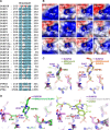
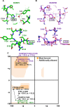
Dual-specificity phosphatases (DUSPs) constitute a subfamily of protein tyrosine phosphatases, and are intimately involved in the regulation of diverse parameters of cellular signaling and essential biological processes. DUSP28 is one of the DUSP subfamily members that is known to be implicated in the progression of hepatocellular and pancreatic cancers, and its biological functions and enzymatic characteristics are mostly unknown. Herein, we present the crystal structure of human DUSP28 determined to 2.1 Å resolution. DUSP28 adopts a typical DUSP fold, which is composed of a central β-sheet covered by α-helices on both sides and contains a well-ordered activation loop, as do other enzymatically active DUSP proteins. The catalytic pocket of DUSP28, however, appears hardly accessible to a substrate because of the presence of nonconserved bulky residues in the protein tyrosine phosphatase signature motif. Accordingly, DUSP28 showed an atypically low phosphatase activity in the biochemical assay, which was remarkably improved by mutations of two nonconserved residues in the activation loop. Overall, this work reports the structural and biochemical basis for understanding a putative oncological therapeutic target, DUSP28, and also provides a unique mechanism for the regulation of enzymatic activity in the DUSP subfamily proteins.
Conflict of interest statement
Figures


Similar articles
-
Role of Conserved Histidine and Serine in the HCXXXXXRS Motif of Human Dual-Specificity Phosphatase 5.J Chem Inf Model. 2019 Apr 22;59(4):1563-1574. doi: 10.1021/acs.jcim.8b00919. Epub 2019 Mar 20. J Chem Inf Model. 2019. PMID: 30835471
-
The family-wide structure and function of human dual-specificity protein phosphatases.Acta Crystallogr D Biol Crystallogr. 2014 Feb;70(Pt 2):421-35. doi: 10.1107/S1399004713029866. Epub 2014 Jan 29. Acta Crystallogr D Biol Crystallogr. 2014. PMID: 24531476
-
High-resolution crystal structure of the catalytic domain of human dual-specificity phosphatase 26.Acta Crystallogr D Biol Crystallogr. 2013 Jun;69(Pt 6):1160-70. doi: 10.1107/S0907444913004770. Epub 2013 May 16. Acta Crystallogr D Biol Crystallogr. 2013. PMID: 23695260
-
Dual Specificity Phosphatase 5-Substrate Interaction: A Mechanistic Perspective.Compr Physiol. 2017 Sep 12;7(4):1449-1461. doi: 10.1002/cphy.c170007. Compr Physiol. 2017. PMID: 28915331 Review.
-
Dual-Specificity Phosphatase Regulation in Neurons and Glial Cells.Int J Mol Sci. 2019 Apr 23;20(8):1999. doi: 10.3390/ijms20081999. Int J Mol Sci. 2019. PMID: 31018603 Free PMC article. Review.
Cited by
-
Structural and biochemical characterization of Siw14: A protein-tyrosine phosphatase fold that metabolizes inositol pyrophosphates.J Biol Chem. 2018 May 4;293(18):6905-6914. doi: 10.1074/jbc.RA117.001670. Epub 2018 Mar 14. J Biol Chem. 2018. PMID: 29540476 Free PMC article.
-
Structural study reveals the temperature-dependent conformational flexibility of Tk-PTP, a protein tyrosine phosphatase from Thermococcus kodakaraensis KOD1.PLoS One. 2018 May 23;13(5):e0197635. doi: 10.1371/journal.pone.0197635. eCollection 2018. PLoS One. 2018. PMID: 29791483 Free PMC article.
-
In Vitro and In Silico Investigation of BCI Anticancer Properties and Its Potential for Chemotherapy-Combined Treatments.Cancers (Basel). 2023 Sep 6;15(18):4442. doi: 10.3390/cancers15184442. Cancers (Basel). 2023. PMID: 37760412 Free PMC article.
References
-
- Tonks NK (2006) Protein tyrosine phosphatases: from genes, to function, to disease. Nat Rev Mol Cell Biol 7: 833–846. doi: 10.1038/nrm2039 - DOI - PubMed
-
- Alonso A, Sasin J, Bottini N, Friedberg I, Friedberg I, Osterman A, et al. (2004) Protein tyrosine phosphatases in the human genome. Cell 117: 699–711. doi: 10.1016/j.cell.2004.05.018 - DOI - PubMed
-
- Patterson KI, Brummer T, O'Brien PM, Daly RJ (2009) Dual-specificity phosphatases: critical regulators with diverse cellular targets. Biochem J 418: 475–489. - PubMed
-
- Rios P, Nunes-Xavier CE, Tabernero L, Kohn M, Pulido R (2014) Dual-specificity phosphatases as molecular targets for inhibition in human disease. Antioxid Redox Signal 20: 2251–2273. doi: 10.1089/ars.2013.5709 - DOI - PubMed
-
- Pulido R, Hooft van Huijsduijnen R (2008) Protein tyrosine phosphatases: dual-specificity phosphatases in health and disease. FEBS J 275: 848–866. doi: 10.1111/j.1742-4658.2008.06250.x - DOI - PubMed
MeSH terms
Substances
LinkOut - more resources
Full Text Sources
Other Literature Sources
Molecular Biology Databases

