Trabecular Bone Morphology Correlates With Skeletal Maturity and Body Composition in Healthy Adolescent Girls
- PMID: 29121215
- PMCID: PMC5761494
- DOI: 10.1210/jc.2017-01785
Trabecular Bone Morphology Correlates With Skeletal Maturity and Body Composition in Healthy Adolescent Girls
Abstract
Context: Growth in healthy children is associated with changes in bone density and microarchitecture. Trabecular morphology is an additional important determinant of bone strength, but little is currently known about trabecular morphology in healthy young people.
Objective: To investigate associations of trabecular morphology with increasing maturity and with body composition in healthy girls.
Design: Cross-sectional study.
Setting: Academic research center.
Participants: Eighty-six healthy girls aged 9 to 18 years.
Main outcome measures: High-resolution peripheral quantitative computed tomography and individual trabecula segmentation were used to assess volumetric bone density, microarchitecture, and trabecular morphology (plate-like vs rod-like) at the distal radius and tibia.
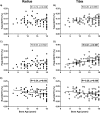
Results: Plate-like bone volume divided by total volume (pBV/TV) increased statistically significantly at the tibia (R = 0.41, P < 0.001), whereas rod-like BV/TV (rBV/TV) decreased statistically significantly at both the radius and tibia (R = -0.34, P = 0.003 and R = -0.28, P = 0.008, respectively) with increasing bone age. In multivariable models, lean mass positively correlated with pBV/TV and plate number at the radius and with plate thickness at both sites. In contrast, fat mass negatively correlated with plate thickness at the tibia and plate surface at both sites. In addition, fat mass positively correlated with rBV/TV and number at the tibia. pBV/TV at both the distal radius and tibia was positively correlated with spine bone mineral density.
Conclusions: Increasing maturity across late childhood and adolescence is associated with changes in trabecular morphology anticipated to contribute to bone strength. Body composition correlates with trabecular morphology, suggesting that muscle mass and adiposity in youth may contribute to long-term skeletal health.
Copyright © 2017 Endocrine Society
Figures


References
-
- Baxter-Jones AD, Faulkner RA, Forwood MR, Mirwald RL, Bailey DA. Bone mineral accrual from 8 to 30 years of age: an estimation of peak bone mass. J Bone Miner Res. 2011;26(8):1729–1739. - PubMed
-
- Seeman E, Hopper JL, Bach LA, Cooper ME, Parkinson E, McKay J, Jerums G. Reduced bone mass in daughters of women with osteoporosis. N Engl J Med. 1989;320(9):554–558. - PubMed
-
- Bouxsein ML, Seeman E. Quantifying the material and structural determinants of bone strength. Best Pract Res Clin Rheumatol. 2009;23(6):741–753. - PubMed
-
- Wang Q, Wang XF, Iuliano-Burns S, Ghasem-Zadeh A, Zebaze R, Seeman E. Rapid growth produces transient cortical weakness: a risk factor for metaphyseal fractures during puberty. J Bone Miner Res. 2010;25(7):1521–1526. - PubMed
Publication types
MeSH terms
Grants and funding
LinkOut - more resources
Full Text Sources
Other Literature Sources
Medical

