

Larval Echinococcus multilocularis infection reduces dextran sulphate sodium-induced colitis in mice by attenuating T helper type 1/type 17-mediated immune reactions
- PMID: 29121394
- PMCID: PMC5904711
- DOI: 10.1111/imm.12860
Larval Echinococcus multilocularis infection reduces dextran sulphate sodium-induced colitis in mice by attenuating T helper type 1/type 17-mediated immune reactions
Abstract
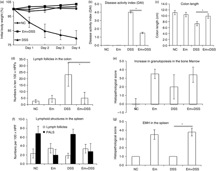
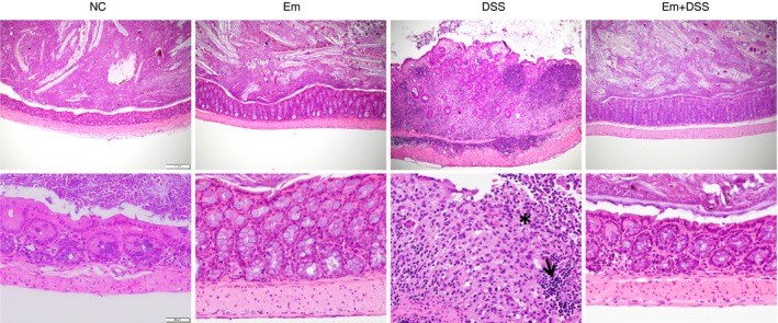
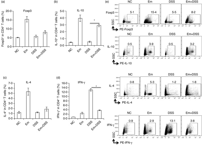
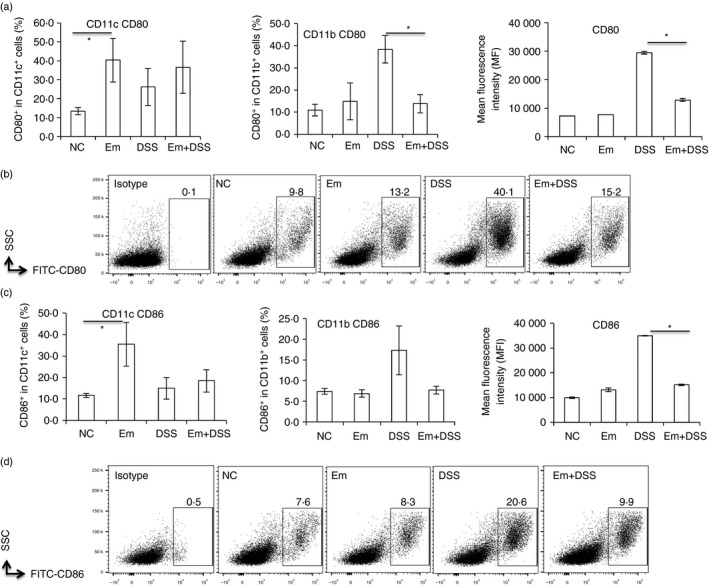
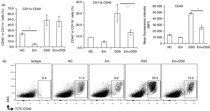
The tumour-like growth of larval Echinococcus multilocularis tissue (causing alveolar echinococcosis, AE) is directly linked to the nature/orientation of the periparasitic host immune-mediated processes. Parasite-mediated immune suppression is a hallmark triggering infection outcome in both chronic human and murine AE. So far, little is known about secondary systemic immune effects of this pathogen on other concomitant diseases, e.g. endogenous gut inflammation. We examined the influence of E. multilocularis infection on murine dextran sodium sulphate (DSS) -induced colitis. At 3 months after E. multilocularis infection (chronic stage), the mice were challenged with 3% DSS in the drinking water for 5 days plus subsequently with tap water (alone) for another 4 days. After necropsy, fixed tissues/organs were sectioned and stained with haematoxylin & eosin for assessing inflammatory reactions. Cytokine levels were measured by flow cytometry and quantitative RT-PCR. Colitis severity was assessed (by board-certified veterinary pathologists) regarding (i) colon length, (ii) weight loss and (iii) a semi-quantitative score of morphological changes. The histopathological analysis of the colon showed a significant reduction of DSS-induced gut inflammation by concomitant E. multilocularis infection, which correlated with down-regulation of T helper type 1 (Th1)/Th17 T-cell responses in the colon tissue. Echinococcus multilocularis infection markedly reduced the severity of DSS-induced gut inflammation upon down-regulation of Th1/Th17 cytokine expression and attenuation of CD11b+ cell activation. In conclusion, E. multilocularis infection remarkably reduces DSS-induced colitis in mice by attenuating Th1/Th17-mediated immune reactions.
Keywords: T helper type 1/type 17 cells; inflammation; parasitic-helminth.
© 2017 The Authors. Immunology Published by John Wiley & Sons Ltd.
Figures

Similar articles
-
Depletion of FoxP3+ Tregs improves control of larval Echinococcus multilocularis infection by promoting co-stimulation and Th1/17 immunity.Immun Inflamm Dis. 2017 Dec;5(4):435-447. doi: 10.1002/iid3.181. Epub 2017 Jun 16. Immun Inflamm Dis. 2017. PMID: 28621034 Free PMC article.
-
Deletion of Fibrinogen-like Protein 2 (FGL-2), a Novel CD4+ CD25+ Treg Effector Molecule, Leads to Improved Control of Echinococcus multilocularis Infection in Mice.PLoS Negl Trop Dis. 2015 May 8;9(5):e0003755. doi: 10.1371/journal.pntd.0003755. eCollection 2015 May. PLoS Negl Trop Dis. 2015. PMID: 25955764 Free PMC article.
-
Berberine ameliorates chronic relapsing dextran sulfate sodium-induced colitis in C57BL/6 mice by suppressing Th17 responses.Pharmacol Res. 2016 Aug;110:227-239. doi: 10.1016/j.phrs.2016.02.010. Epub 2016 Mar 9. Pharmacol Res. 2016. PMID: 26969793
-
Molecular survival strategies of Echinococcus multilocularis in the murine host.Parasitol Int. 2006;55 Suppl:S45-9. doi: 10.1016/j.parint.2005.11.006. Epub 2005 Dec 13. Parasitol Int. 2006. PMID: 16352460 Review.
-
Immunoregulation in larval Echinococcus multilocularis infection.Parasite Immunol. 2016 Mar;38(3):182-92. doi: 10.1111/pim.12292. Parasite Immunol. 2016. PMID: 26536823 Review.
Cited by
-
An exploration of alginate oligosaccharides modulating intestinal inflammatory networks via gut microbiota.Front Microbiol. 2023 Jan 26;14:1072151. doi: 10.3389/fmicb.2023.1072151. eCollection 2023. Front Microbiol. 2023. PMID: 36778853 Free PMC article. Review.
-
Artemisia argyi extract alleviates inflammation in a DSS-induced colitis mouse model and enhances immunomodulatory effects in lymphoid tissues.BMC Complement Med Ther. 2022 Mar 11;22(1):64. doi: 10.1186/s12906-022-03536-x. BMC Complement Med Ther. 2022. PMID: 35277165 Free PMC article.
-
Effects of maternal Echinococcus multilocularis infection on colitis susceptibility and gut microbiota of offspring.Parasit Vectors. 2025 Jul 26;18(1):299. doi: 10.1186/s13071-025-06915-8. Parasit Vectors. 2025. PMID: 40713878 Free PMC article.
-
Schistosoma japonicum peptide SJMHE1 inhibits acute and chronic colitis induced by dextran sulfate sodium in mice.Parasit Vectors. 2021 Sep 6;14(1):455. doi: 10.1186/s13071-021-04977-y. Parasit Vectors. 2021. PMID: 34488863 Free PMC article.
-
Echinococcus multilocularis protoscoleces enhance glycolysis to promote M2 Macrophages through PI3K/Akt/mTOR Signaling Pathway.Pathog Glob Health. 2023 Jun;117(4):409-416. doi: 10.1080/20477724.2022.2104055. Epub 2022 Jul 25. Pathog Glob Health. 2023. PMID: 35876088 Free PMC article.
References
-
- Vuitton DA. The ambiguous role of immunity in echinococcosis: protection of the host or of the parasite? Acta Trop 2003; 85:119–32. - PubMed
-
- Vuitton DA, Zhang SL, Yang Y, Godot V, Beurton I, Mantion G et al Survival strategy of Echinococcus multilocularis in the human host. Parasitol Int 2006; 55(Suppl):S51–5. - PubMed
-
- Gottstein B, Wang J, Boubaker G, Marinova I, Spiliotis M, Muller N et al Susceptibility versus resistance in alveolar echinococcosis (larval infection with Echinococcus multilocularis). Vet Parasitol 2015; 213:103–9. - PubMed
-
- Wang J, Gottstein B. Immunoregulation in larval Echinococcus multilocularis infection. Parasite Immunol 2016; 38:182–92. - PubMed
Publication types
MeSH terms
Substances
Supplementary concepts
LinkOut - more resources
Full Text Sources
Other Literature Sources
Research Materials
Miscellaneous
