Schwann cells participate in synapse elimination at the developing neuromuscular junction
- PMID: 29121585
- PMCID: PMC5732880
- DOI: 10.1016/j.conb.2017.10.010
Schwann cells participate in synapse elimination at the developing neuromuscular junction
Abstract
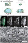
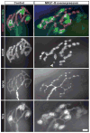
During the initial stages of innervation of developing skeletal muscles, the terminal branches of axons from multiple motor neurons form neuromuscular junctions (NMJs) on a small region of each muscle fiber, the motor endplate. Subsequently, the number of axonal inputs at the endplate region is reduced so that, at maturity, each muscle fiber is innervated by the terminals of a single motor neuron. The Schwann cells associated with the axon terminals are involved in the removal of these synapses but do not select the axon that is ultimately retained on each fiber. Schwann cells perform this function by disconnecting terminal branches from the myofiber surface and by attacking them phagocytically. Here we discuss how this behavior is regulated and argue that such regulation is not unique to development of neuromuscular innervation but is also expressed in the response of the mature NMJ to various manipulations and pathologies.
Copyright © 2017 Elsevier Ltd. All rights reserved.
Conflict of interest statement
The authors declare no conflict of interest.
Figures


Similar articles
-
Neuregulin1 displayed on motor axons regulates terminal Schwann cell-mediated synapse elimination at developing neuromuscular junctions.Proc Natl Acad Sci U S A. 2016 Jan 26;113(4):E479-87. doi: 10.1073/pnas.1519156113. Epub 2016 Jan 11. Proc Natl Acad Sci U S A. 2016. PMID: 26755586 Free PMC article.
-
Neuron-glia interactions: the roles of Schwann cells in neuromuscular synapse formation and function.Biosci Rep. 2011 Oct;31(5):295-302. doi: 10.1042/BSR20100107. Biosci Rep. 2011. PMID: 21517783 Free PMC article. Review.
-
Loss of glial neurofascin155 delays developmental synapse elimination at the neuromuscular junction.J Neurosci. 2014 Sep 17;34(38):12904-18. doi: 10.1523/JNEUROSCI.1725-14.2014. J Neurosci. 2014. PMID: 25232125 Free PMC article.
-
Blocking skeletal muscle DHPRs/Ryr1 prevents neuromuscular synapse loss in mutant mice deficient in type III Neuregulin 1 (CRD-Nrg1).PLoS Genet. 2019 Mar 14;15(3):e1007857. doi: 10.1371/journal.pgen.1007857. eCollection 2019 Mar. PLoS Genet. 2019. PMID: 30870432 Free PMC article.
-
Perisynaptic schwann cells - The multitasking cells at the developing neuromuscular junctions.Semin Cell Dev Biol. 2020 Aug;104:31-38. doi: 10.1016/j.semcdb.2020.02.011. Epub 2020 Mar 5. Semin Cell Dev Biol. 2020. PMID: 32147379 Review.
Cited by
-
Ablation of Lrp4 in Schwann Cells Promotes Peripheral Nerve Regeneration in Mice.Biology (Basel). 2021 May 21;10(6):452. doi: 10.3390/biology10060452. Biology (Basel). 2021. PMID: 34063992 Free PMC article.
-
Wesley J. Thompson (1947-2019).Front Mol Neurosci. 2020 Jun 12;13:91. doi: 10.3389/fnmol.2020.00091. eCollection 2020. Front Mol Neurosci. 2020. PMID: 32595450 Free PMC article. No abstract available.
-
A Novel Optical Tissue Clearing Protocol for Mouse Skeletal Muscle to Visualize Endplates in Their Tissue Context.Front Cell Neurosci. 2019 Feb 27;13:49. doi: 10.3389/fncel.2019.00049. eCollection 2019. Front Cell Neurosci. 2019. PMID: 30873005 Free PMC article.
-
Developmental neuromuscular synapse elimination: Activity-dependence and potential downstream effector mechanisms.Neurosci Lett. 2020 Jan 23;718:134724. doi: 10.1016/j.neulet.2019.134724. Epub 2019 Dec 23. Neurosci Lett. 2020. PMID: 31877335 Free PMC article. Review.
-
Adenosine Receptors in Developing and Adult Mouse Neuromuscular Junctions and Functional Links With Other Metabotropic Receptor Pathways.Front Pharmacol. 2018 Apr 24;9:397. doi: 10.3389/fphar.2018.00397. eCollection 2018. Front Pharmacol. 2018. PMID: 29740322 Free PMC article.
References
-
- Tello F. Degeneration et regeneration des plagues motrices. Travaux du Laboratorire de Recherches Biologiques de l’Universit’e de Madrid. 1907;5:117–149.
-
- Walsh MK, Lichtman JW. In vivo time-lapse imaging of synaptic takeover associated with naturally occurring synapse elimination. Neuron. 2003;37:67–73. The authors utilized transgenic expression of two distinct fluorochromes that by variegation mark separate pool of axons innervating muscle fibers and examine vitally the competition between convergent axons during the last stages of synapse elimination. - PubMed
-
- Turney SG, Lichtman JW. Reversing the outcome of synapse elimination at developing neuromuscular junctions in vivo: evidence for synaptic competition and its mechanism. PLoS Biol. 2012;10:e1001352. The investigators utilized laser ablation of axons to manipulate the innervation of individual NMJs during the synapse elimination and demonstrated that the winner of the interneuronal competition at individual junction is not predetermined. - PMC - PubMed
Publication types
MeSH terms
Grants and funding
LinkOut - more resources
Full Text Sources
Other Literature Sources

