Comparative Study
. 2017 Nov 9;12(11):e0187948.
doi: 10.1371/journal.pone.0187948. eCollection 2017.

Hypertrophic cardiomyopathy clinical phenotype is independent of gene mutation and mutation dosage

Affiliations
Comparative Study

Hypertrophic cardiomyopathy clinical phenotype is independent of gene mutation and mutation dosage

Shiv Kumar Viswanathan et al. PLoS One. .

Abstract

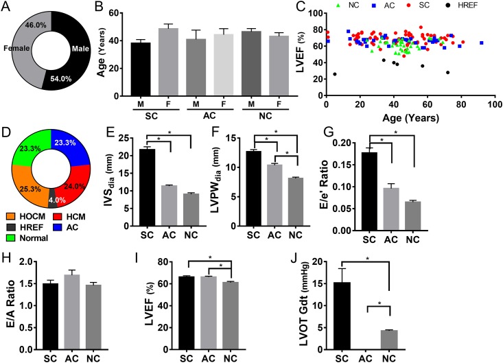
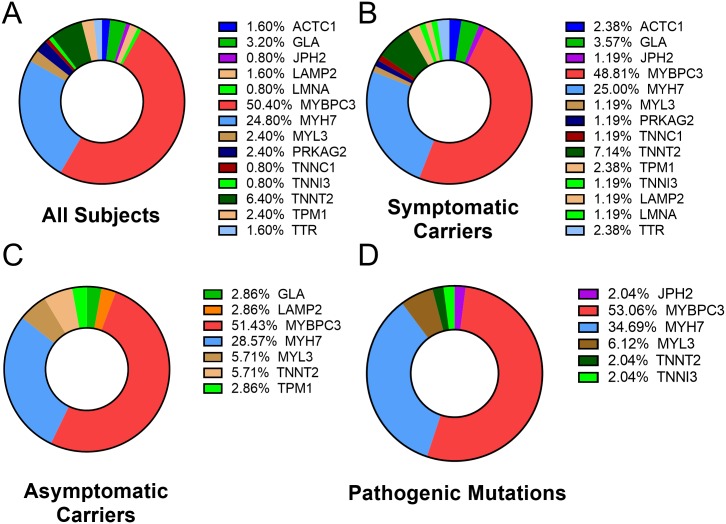
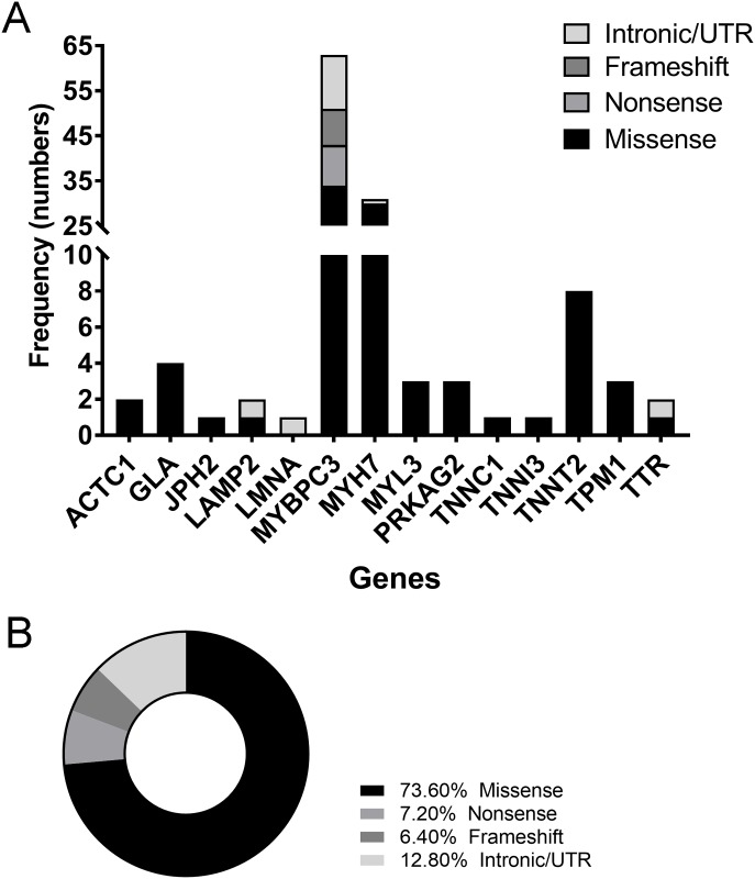
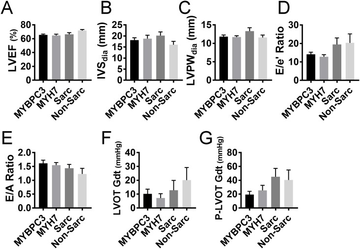
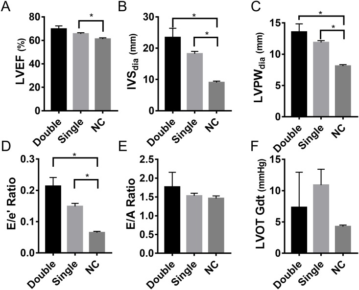
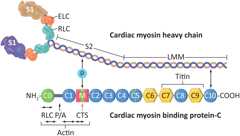
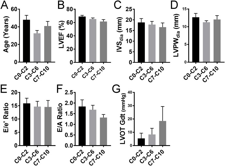
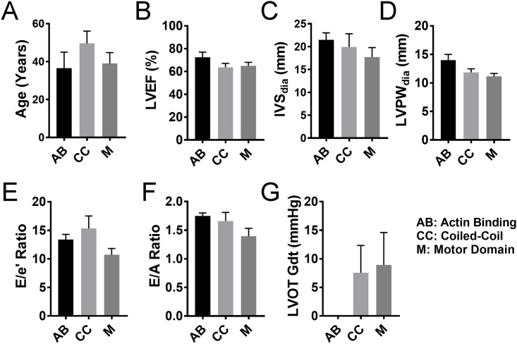
Over 1,500 gene mutations are known to cause hypertrophic cardiomyopathy (HCM). Previous studies suggest that cardiac β-myosin heavy chain (MYH7) gene mutations are commonly associated with a more severe phenotype, compared to cardiac myosin binding protein-C (MYBPC3) gene mutations with milder phenotype, incomplete penetrance and later age of onset. Compound mutations can worsen the phenotype. This study aimed to validate these comparative differences in a large cohort of individuals and families with HCM. We performed genome-phenome correlation among 80 symptomatic HCM patients, 35 asymptomatic carriers and 35 non-carriers, using an 18-gene clinical diagnostic HCM panel. A total of 125 mutations were identified in 14 genes. MYBPC3 and MYH7 mutations contributed to 50.0% and 24.4% of the HCM patients, respectively, suggesting that MYBPC3 mutations were the most frequent cause of HCM in our cohort. Double mutations were found in only nine HCM patients (7.8%) who were phenotypically indistinguishable from single-mutation carriers. Comparisons of clinical parameters of MYBPC3 and MYH7 mutants were not statistically significant, but asymptomatic carriers had high left ventricular ejection fraction and diastolic dysfunction when compared to non-carriers. The presence of double mutations increases the risk for symptomatic HCM with no change in severity, as determined in this study subset. The pathologic effects of MYBPC3 and MYH7 were found to be independent of gene mutation location. Furthermore, HCM pathology is independent of protein domain disruption in both MYBPC3 and MYH7. These data provide evidence that MYBPC3 mutations constitute the preeminent cause of HCM and that they are phenotypically indistinguishable from HCM caused by MYH7 mutations.


Conflict of interest statement

Competing Interests: The authors have declared that no competing interests exist.

Figures

Fig 1
Fig 1. Study subject characteristics across three cohorts and clinical evaluation profile.
A, Distribution of males and females in the study population represented as percentages. B, Age and gender distribution among the three cohorts, probands with definitive HCM/HOCM or dHCM clinical phenotype (symptomatic carriers, SC), family members with no HCM phenotype (asymptomatic carriers, AC), and non-carriers (NC) unrelated community-based population confirmed by echocardiography to have normal cardiac structure and function. Data are represented as means with standard error of mean (SEM) C, Age-based distribution of left ventricular ejection fraction across the three cohorts; red markers indicate HCM, blue markers indicate AC, and green markers indicate NC. Black markers indicate dHCM subjects with ejection fraction ≤45%. D, Diagnostic subclassification of subjects with gene mutation represented as a percentage of total. E-J panels show various echocardiographic parameters that reflect HCM severity presented as mean with standard error of mean. IVS, interventricular septum thickness (E); LVPW, left ventricular posterior wall (free wall) thickness (F); E/e’ ratio, ratio of early transmitral flow (E) to left ventricular early diastolic velocity (e’) (G); E/A ratio, ratio of early (E) to late (A) ventricular filling velocities (H); LVEF, left ventricular ejection fraction (I); LVOT-Gdt, LV outflow tract peak gradient (J). * indicates p value of 0.01 or lower by one-way ANOVA analysis.
Fig 2
Fig 2. Genetic evaluation profile of the study cohort.
A-C, show the percentage of gene mutation contribution among all subjects (A); HCM (B); and AC (C). Pathogenic mutation distribution among the various causative genes (D). MYBPC3 and MYH7 mutations are causative in >80% of all subjects, irrespective of their current phenotypic pathogenicity.
Fig 3
Fig 3. Frequency distribution of mutations in HCM-causing genes.
The mutation distribution in 13 of the 18 HCM causative genes based on mutation class, including missense, nonsense, frameshift mutations and intronic and UTR variations (A). Distribution of mutation types among the causative genes (B), with missense and nonsense mutation contributing more than 80% of all classes of mutations.
Fig 4
Fig 4. Phenotypic effect of mutations in MYBPC3, MYH7, sarcomeric (Sarc) and non-sarcomeric (Non-Sarc) genes.
The severity of pathogenesis, as measured by echocardiography (A-G). Echo parameters that reflect HCM severity include LVEF, left ventricular ejection fraction (A); IVS, interventricular septum thickness (B); LVPW, left ventricular posterior wall (free wall) thickness (C); E/e’ ratio, ratio of early transmitral flow (E) to left ventricular early diastolic velocity (e’) (D); E/A ratio, ratio of early (E) to late (A) ventricular filling velocities (E); LVOT-Gdt, LV outflow tract peak gradient (F) and provoked LVOT gradient (P-LVOT Gdt) (G). All parameters are represented as mean with SEM.
Fig 5
Fig 5. Effect of gene mutation dosage on the pathogenesis of HCM.
Comparison of double mutation (Dbl) and single mutation (Sing) with non-carrier (NC) subjects. The severity of pathogenesis between the groups was measured by LVEF, left ventricular ejection fraction (A); IVS, interventricular septum thickness (B); LVPW, left ventricular posterior wall (free wall) thickness (C); E/e’ ratio, ratio of early transmitral flow (E) to left ventricular early diastolic velocity (e’) (D); E/A ratio, ratio of early (E) to late (A) ventricular filling velocities (E) and LVOT-Gdt, LV outflow tract peak gradient (F). * indicates p value of 0.01 or lower by one-way ANOVA analysis.
Fig 6
Fig 6. Schematic diagram of cardiac β-myosin heavy chain (β-MHC) and cardiac myosin binding protein-C (cMyBP-C) proteins.
cMyBP-C shows the C0-C10 domains and regions of interaction with other proteins, including β-myosin. S1, Myosin heads; ELC, essential light chain; RLC, regulatory light chain; S2, myosin neck region; LMM, light meromyosin. P/A, Proline- and Alanine-rich domain. CTS, Calpain-targeted site.
Fig 7
Fig 7. Mutations in cMyBP-C protein interaction domains and their effect on the pathogenesis of HCM.
C0-C10 indicates the 11 domains in cMyBP-C protein. The severity of pathogenesis among the groups (N’ C0-C2, C3-C6 and C’ C7-C10 domains) was measured by age at evaluation. (A), LVEF, left ventricular ejection fraction (B); IVS, interventricular septum thickness (C); LVPW, left ventricular posterior wall (free wall) thickness (D); E/e’ ratio, ratio of early transmitral flow (E) to left ventricular early diastolic velocity (e’) (E); E/A ratio, ratio of early (E) to late (A) ventricular filling velocities (F) and LVOT-Gdt, LV outflow tract peak gradient (G); n = 14 for C0-C2, n = 19 for C3-C6 and n = 17 for C7-C10.
Fig 8
Fig 8. Mutations in β-MHC interaction domains and their effect on the pathogenesis of HCM.
The three domains of β-MHC, namely the AB, Actin binding domain; CC, Coiled-coil domain; and M, Motor domain, were compared to each other in order to assess the pathogenicity of mutations that occur in these domains. The severity of pathogenesis among the groups was measured by age at evaluation. (A), LVEF, left ventricular ejection fraction (B); IVS, interventricular septum thickness (C); LVPW, left ventricular posterior wall (free wall) thickness (D); E/e’ ratio, ratio of early transmitral flow (E) to left ventricular early diastolic velocity (e’) (E); E/A ratio, ratio of early (E) to late (A) ventricular filling velocities (F) and LVOT-Gdt, LV outflow tract peak gradient (G); n = 4 for AB, n = 13 for CC and n = 13 for M.

Similar articles

Cited by

References

    1. Semsarian C, Ingles J, Maron MS, Maron BJ. New perspectives on the prevalence of hypertrophic cardiomyopathy. J Am Coll Cardiol. 2015;65(12):1249–54. doi: 10.1016/j.jacc.2015.01.019 . - DOI - PubMed
    1. Authors/Task Force m, Elliott PM, Anastasakis A, Borger MA, Borggrefe M, Cecchi F, et al. 2014 ESC Guidelines on diagnosis and management of hypertrophic cardiomyopathy: the Task Force for the Diagnosis and Management of Hypertrophic Cardiomyopathy of the European Society of Cardiology (ESC). Eur Heart J. 2014;35(39):2733–79. doi: 10.1093/eurheartj/ehu284 . - DOI - PubMed
    1. Varnava AM, Elliott PM, Mahon N, Davies MJ, McKenna WJ. Relation between myocyte disarray and outcome in hypertrophic cardiomyopathy. Am J Cardiol. 2001;88(3):275–9. . - PubMed
    1. O'Mahony C, Jichi F, Pavlou M, Monserrat L, Anastasakis A, Rapezzi C, et al. A novel clinical risk prediction model for sudden cardiac death in hypertrophic cardiomyopathy (HCM risk-SCD). Eur Heart J. 2014;35(30):2010–20. doi: 10.1093/eurheartj/eht439 . - DOI - PubMed
    1. Ullal AJ, Abdelfattah RS, Ashley EA, Froelicher VF. Hypertrophic Cardiomyopathy as a Cause of Sudden Cardiac Death in the Young: A Meta-Analysis. Am J Med. 2016;129(5):486–96 e2. doi: 10.1016/j.amjmed.2015.12.027 . - DOI - PubMed

Publication types

LinkOut - more resources