

Apoptosis inhibitor of macrophage depletion decreased M1 macrophage accumulation and the incidence of cardiac rupture after myocardial infarction in mice
- PMID: 29121663
- PMCID: PMC5679665
- DOI: 10.1371/journal.pone.0187894
Apoptosis inhibitor of macrophage depletion decreased M1 macrophage accumulation and the incidence of cardiac rupture after myocardial infarction in mice
Abstract
Background: Cardiac rupture is an important cause of death in the acute phase after myocardial infarction (MI). Macrophages play a pivotal role in cardiac remodeling after MI. Apoptosis inhibitor of macrophage (AIM) is secreted specifically by macrophages and contributes to macrophage accumulation in inflamed tissue by maintaining survival and recruiting macrophages. In this study, we evaluated the role of AIM in macrophage accumulation in the infarcted myocardium and cardiac rupture after MI.
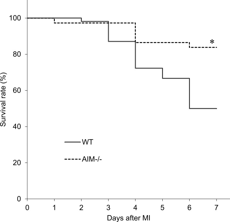
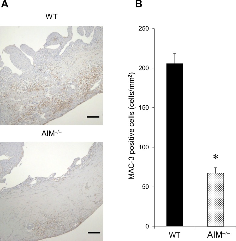
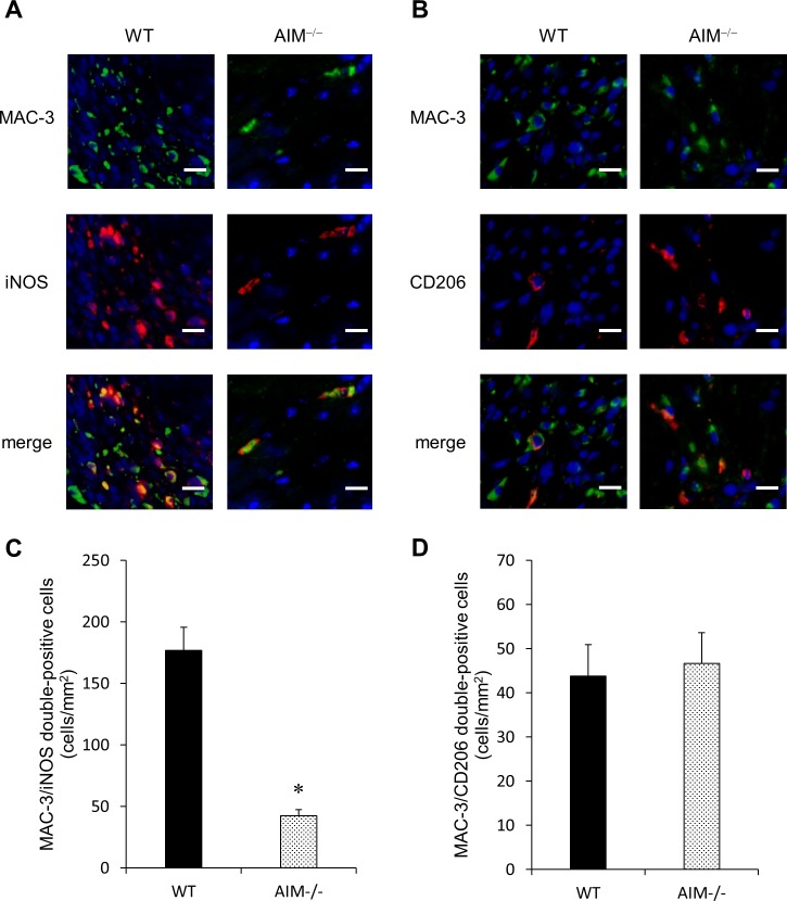
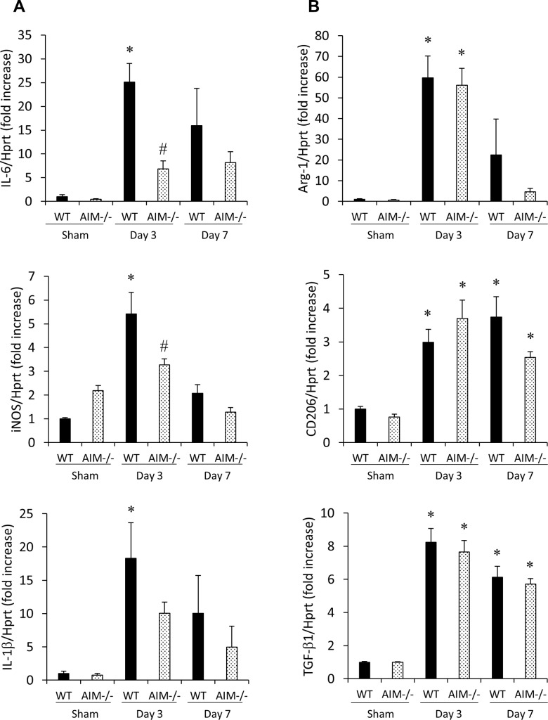
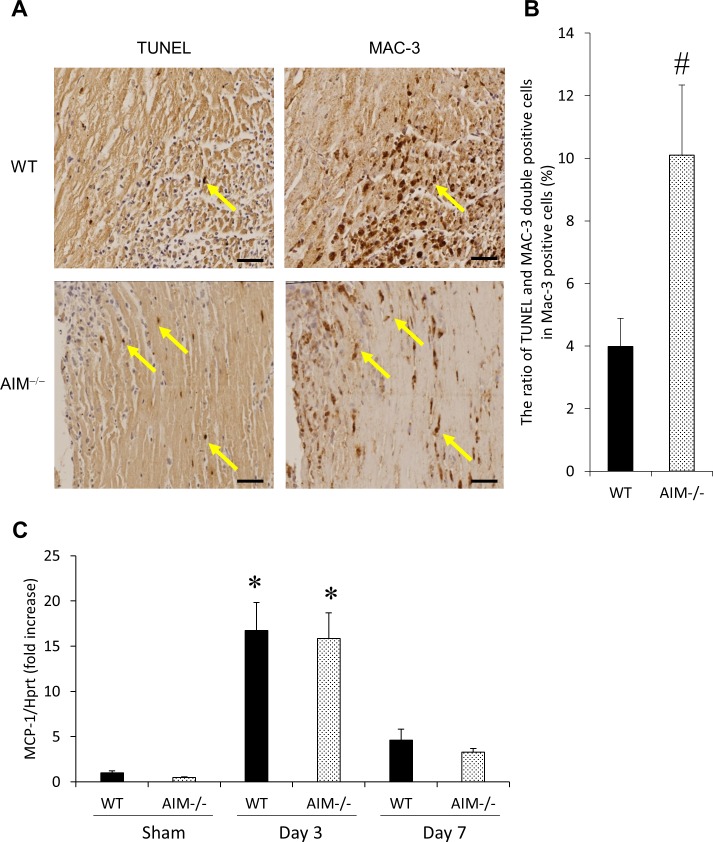
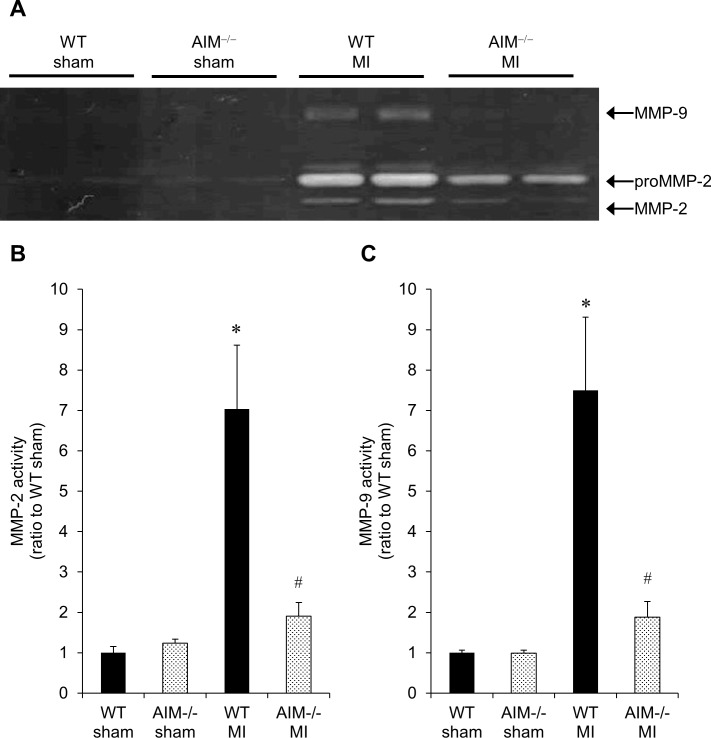
Methods and results: Wild-type (WT) and AIM‒/‒ mice underwent permanent left coronary artery ligation and were followed-up for 7 days. Macrophage accumulation and phenotypes (M1 pro-inflammatory macrophage or M2 anti-inflammatory macrophage) were evaluated by immunohistological analysis and RT-PCR. Matrix metalloproteinase (MMP) activity levels were measured by gelatin zymography. The survival rate was significantly higher (81.1% vs. 48.2%, P<0.05), and the cardiac rupture rate was significantly lower in AIM‒/‒ mice than in WT mice (10.8% vs. 31.5%, P<0.05). The number of M1 macrophages and the expression levels of M1 markers (iNOS and IL-6) in the infarcted myocardium were significantly lower in AIM‒/‒ mice than in WT mice. In contrast, there was no difference in the number of M2 macrophages and the expression of M2 markers (Arg-1, CD206 and TGF-β1) between the two groups. The ratio of apoptotic macrophages in the total macrophages was significantly higher in AIM‒/‒ mice than in WT mice, although MCP-1 expression did not differ between the two groups. MMP-2 and 9 activity levels in the infarcted myocardium were significantly lower in AIM‒/‒ mice than in WT mice.
Conclusions: These findings suggest that AIM depletion decreases the levels of M1 macrophages, which are a potent source of MMP-2 and 9, in the infarcted myocardium in the acute phase after MI by promoting macrophage apoptosis, and leads to a decrease in the incidence of cardiac rupture and improvements in survival rates.
Conflict of interest statement
Figures

Similar articles
-
Targeted deletion of class A macrophage scavenger receptor increases the risk of cardiac rupture after experimental myocardial infarction.Circulation. 2007 Apr 10;115(14):1904-11. doi: 10.1161/CIRCULATIONAHA.106.671198. Epub 2007 Mar 26. Circulation. 2007. PMID: 17389263
-
Calpastatin overexpression impairs postinfarct scar healing in mice by compromising reparative immune cell recruitment and activation.Am J Physiol Heart Circ Physiol. 2015 Dec 1;309(11):H1883-93. doi: 10.1152/ajpheart.00594.2015. Epub 2015 Oct 9. Am J Physiol Heart Circ Physiol. 2015. PMID: 26453333
-
Targeted deletion or pharmacological inhibition of MMP-2 prevents cardiac rupture after myocardial infarction in mice.J Clin Invest. 2005 Mar;115(3):599-609. doi: 10.1172/JCI22304. J Clin Invest. 2005. PMID: 15711638 Free PMC article.
-
The Protective Role of TREM2 in the Heterogenous Population of Macrophages during Post-Myocardial Infarction Inflammation.Int J Mol Sci. 2023 Mar 14;24(6):5556. doi: 10.3390/ijms24065556. Int J Mol Sci. 2023. PMID: 36982629 Free PMC article. Review.
-
Knowledge gaps to understanding cardiac macrophage polarization following myocardial infarction.Biochim Biophys Acta. 2016 Dec;1862(12):2288-2292. doi: 10.1016/j.bbadis.2016.05.013. Epub 2016 May 27. Biochim Biophys Acta. 2016. PMID: 27240543 Free PMC article. Review.
Cited by
-
Human umbilical cord mesenchymal stem cells alleviate ongoing autoimmune dacryoadenitis in rabbits via polarizing macrophages into an anti-inflammatory phenotype.Exp Eye Res. 2020 Feb;191:107905. doi: 10.1016/j.exer.2019.107905. Epub 2019 Dec 28. Exp Eye Res. 2020. PMID: 31891674 Free PMC article.
-
Macrophage plasticity: signaling pathways, tissue repair, and regeneration.MedComm (2020). 2024 Aug 1;5(8):e658. doi: 10.1002/mco2.658. eCollection 2024 Aug. MedComm (2020). 2024. PMID: 39092292 Free PMC article. Review.
-
The comprehensive role of apoptosis inhibitor of macrophage (AIM) in pathological conditions.Clin Exp Immunol. 2023 Jun 5;212(3):184-198. doi: 10.1093/cei/uxac095. Clin Exp Immunol. 2023. PMID: 36427004 Free PMC article. Review.
-
Macrophage CARD9 mediates cardiac injury following myocardial infarction through regulation of lipocalin 2 expression.Signal Transduct Target Ther. 2023 Oct 13;8(1):394. doi: 10.1038/s41392-023-01635-w. Signal Transduct Target Ther. 2023. PMID: 37828006 Free PMC article.
-
Cardiac Myocyte-Specific Overexpression of FASTKD1 Prevents Ventricular Rupture After Myocardial Infarction.J Am Heart Assoc. 2023 Feb 21;12(4):e025867. doi: 10.1161/JAHA.122.025867. Epub 2023 Feb 15. J Am Heart Assoc. 2023. PMID: 36789858 Free PMC article.
References
-
- Reed GW, Rossi J rey E, Cannon CP. Acute myocardial infarction. Lancet. 2016;16: 30677–8. doi: 10.1016/j.disamonth.2012.12.004 - DOI - PubMed
-
- Nichols M, Townsend N, Scarborough P, Rayner M, Lozano R, Naghavi M, et al. Cardiovascular disease in Europe 2014: epidemiological update. Eur Heart J. 2014;35: 2950–9. doi: 10.1093/eurheartj/ehu299 - DOI - PubMed
-
- Toru Takii, Satoshi Yasuda, Takahashi Jun, Kenta Ito, Nobuyuki Shiba, Shirato Kunio SH. Trends in Acute Myocardial Infarction Incidence and Mortality over 30 years in Japan. Circ J. 2010;74: 93–100. - PubMed
-
- Yip H, Wu C, Chang H, Wang C-P, Cheng C-I, Chua S, et al. Cardiac rupture complicating acute myocardial infarction in the direct percutaneous coronary intervention reperfusion era. Chest. 2003;124: 565–71. doi: 10.1378/chest.124.2.565 - DOI - PubMed
-
- Honda S, Asaumi Y, Yamane T, Nagai T, Miyagi T, Noguchi T, et al. Trends in the Clinical and Pathological Characteristics of Cardiac Rupture in Patients With Acute Myocardial Infarction Over 35 Years. J Am Hear Assoc. 2014;3: e000984 doi: 10.1161/JAHA.114.000984 - DOI - PMC - PubMed
MeSH terms
Substances
LinkOut - more resources
Full Text Sources
Other Literature Sources
Medical
Molecular Biology Databases
Research Materials
Miscellaneous
