

Heart rate and blood pressure changes during sleep-waking cycles and cataplexy in narcoleptic dogs
- PMID: 2912173
- PMCID: PMC9050242
- DOI: 10.1152/ajpheart.1989.256.1.H111
Heart rate and blood pressure changes during sleep-waking cycles and cataplexy in narcoleptic dogs
Abstract
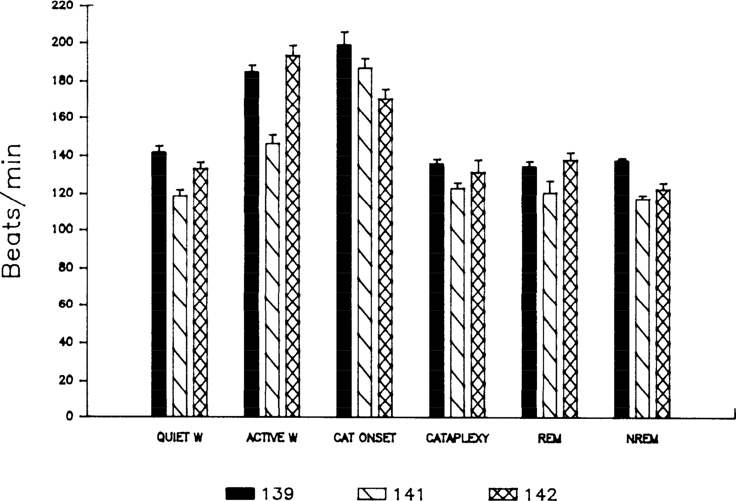
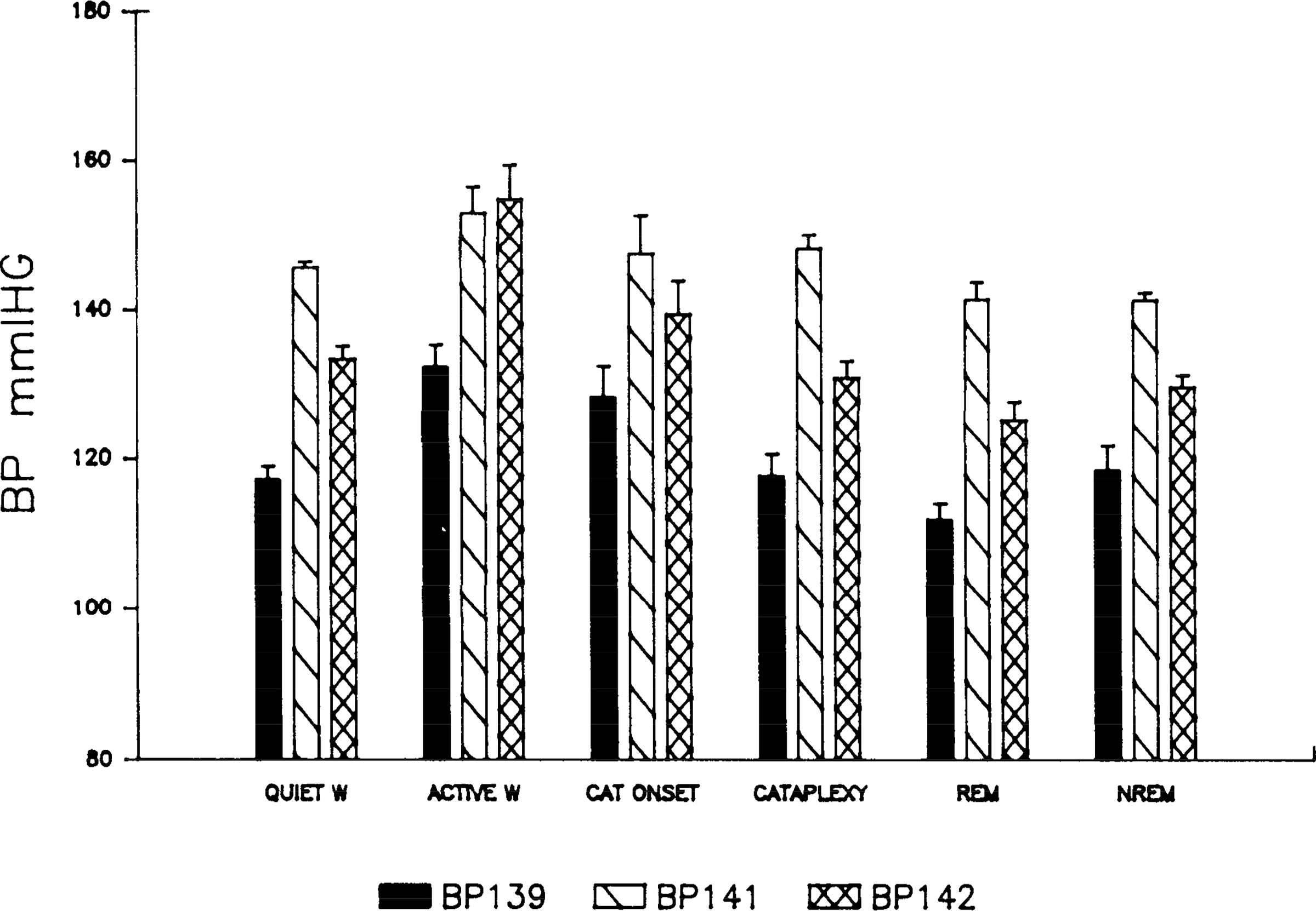
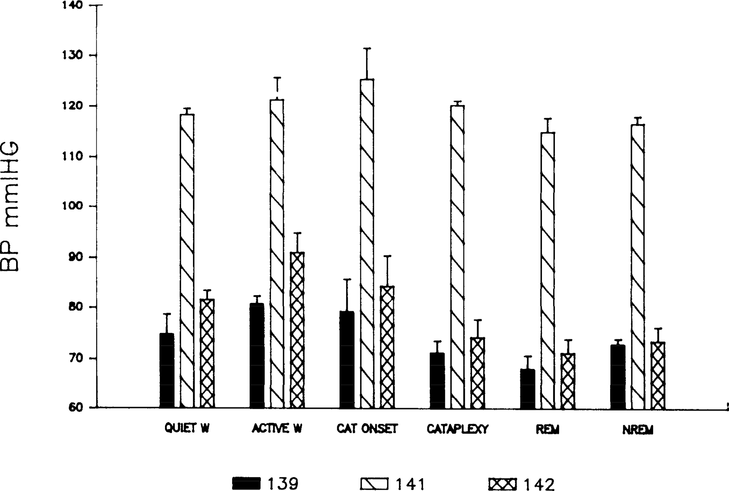
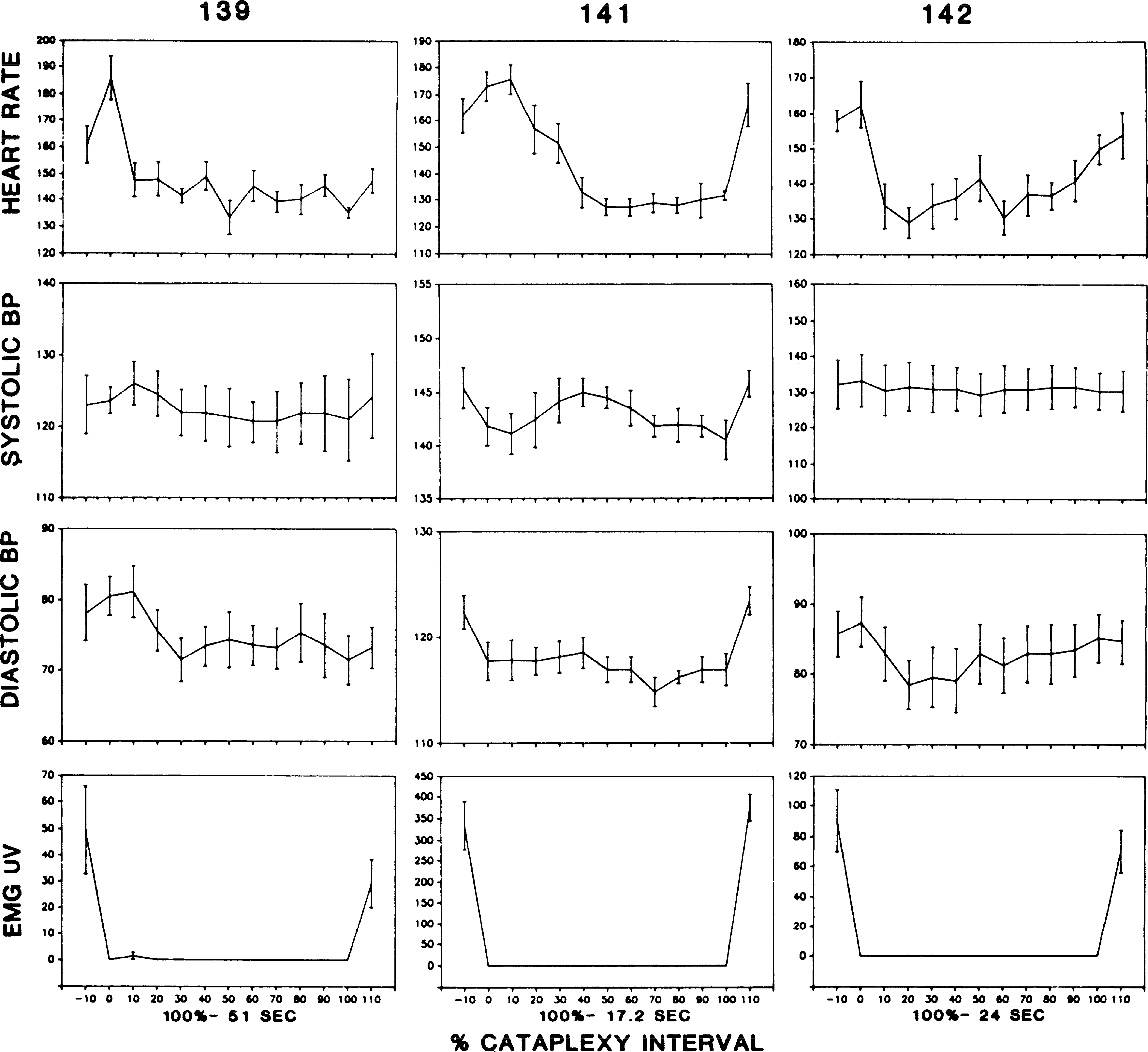
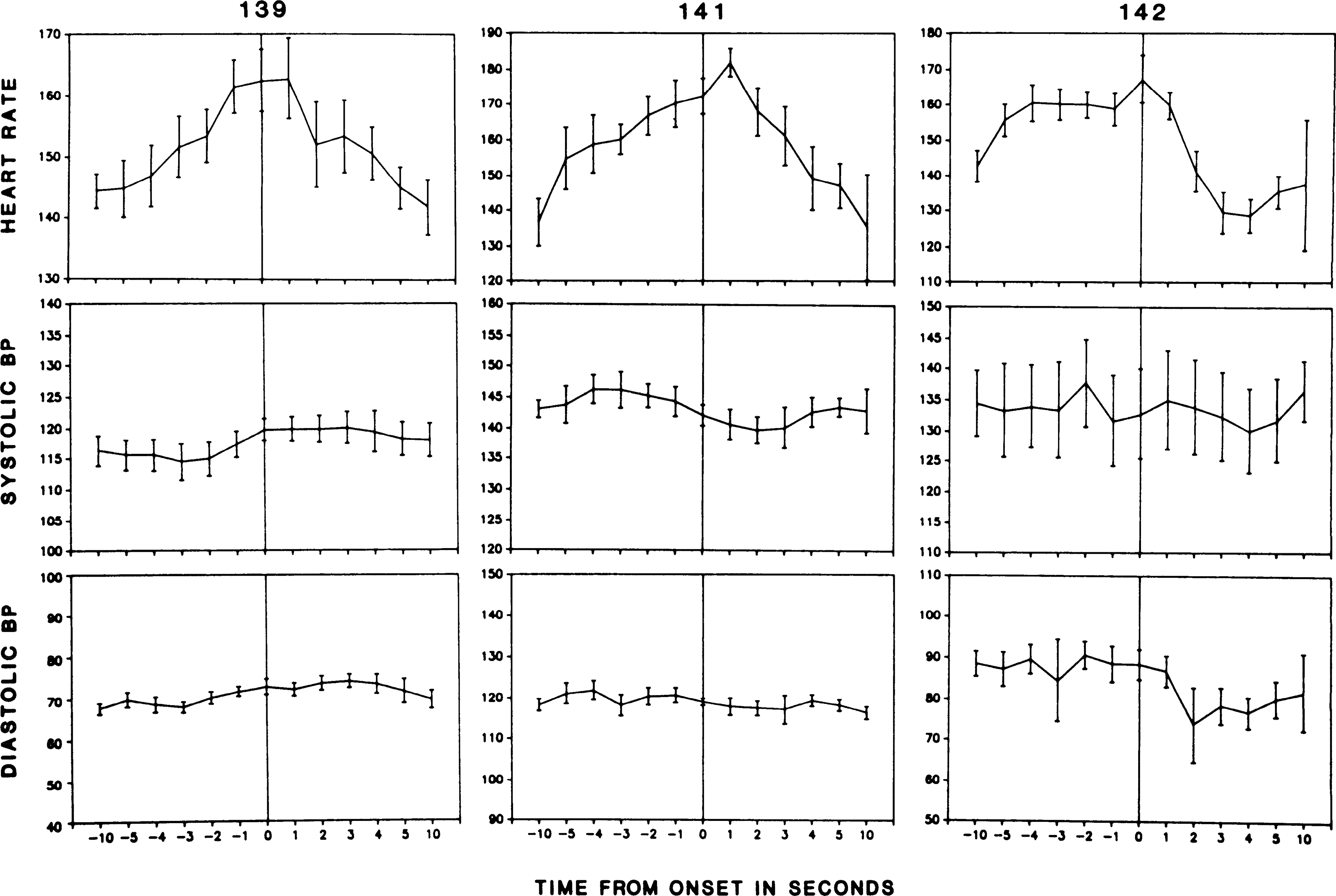
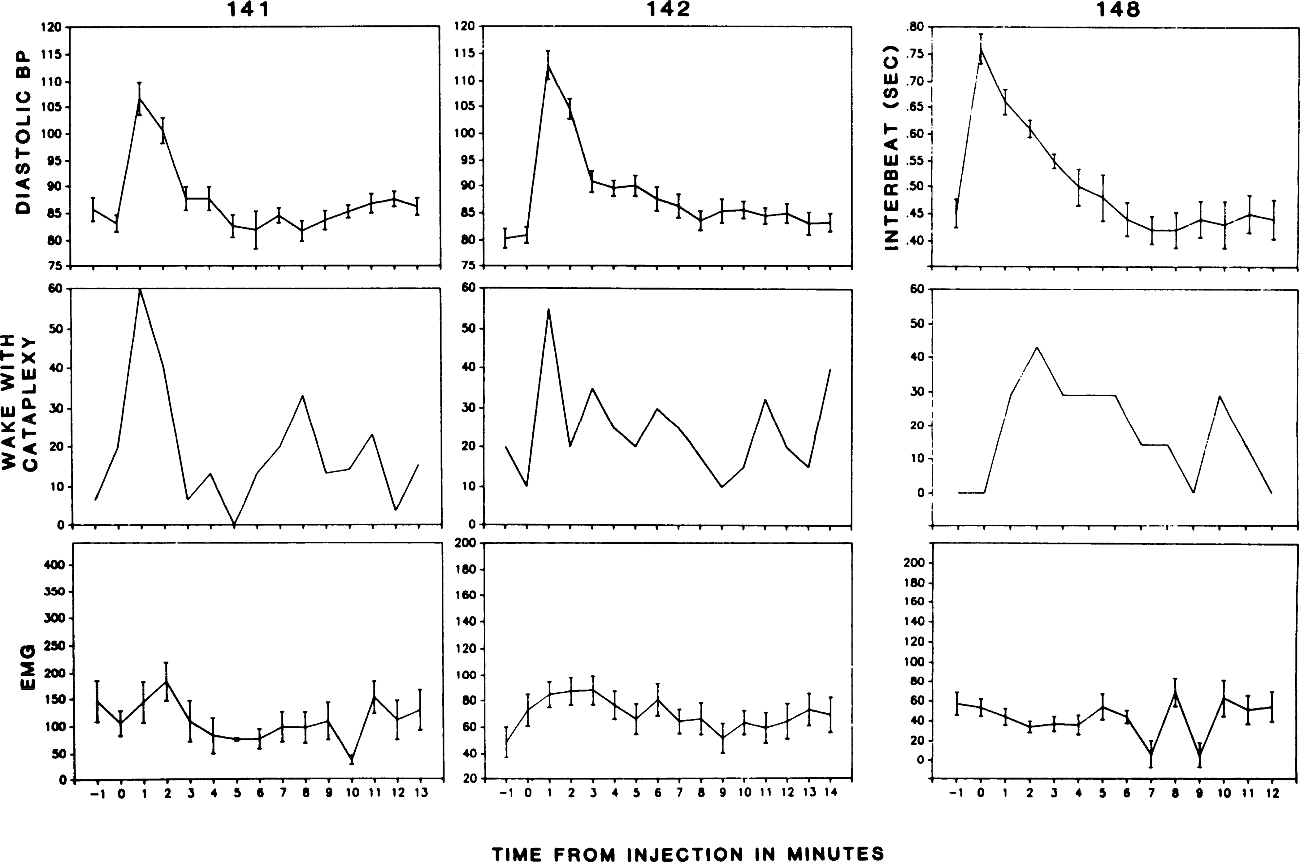
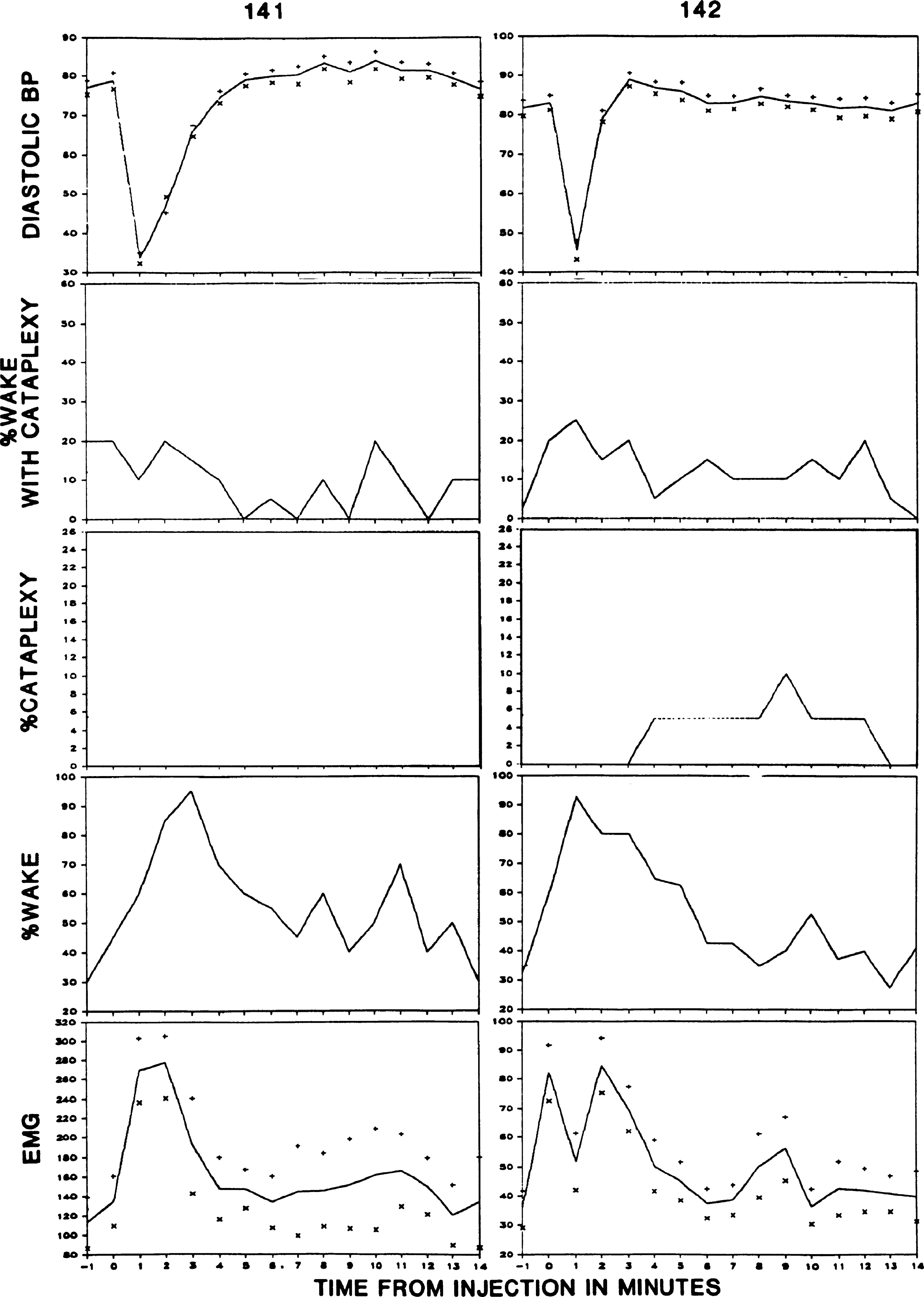
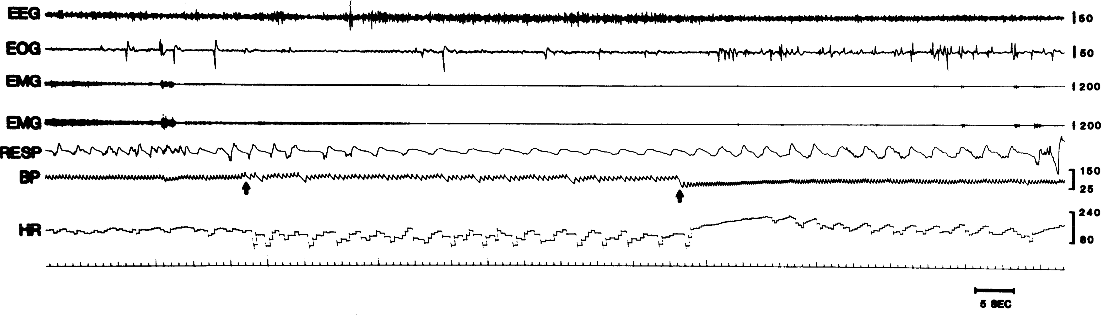
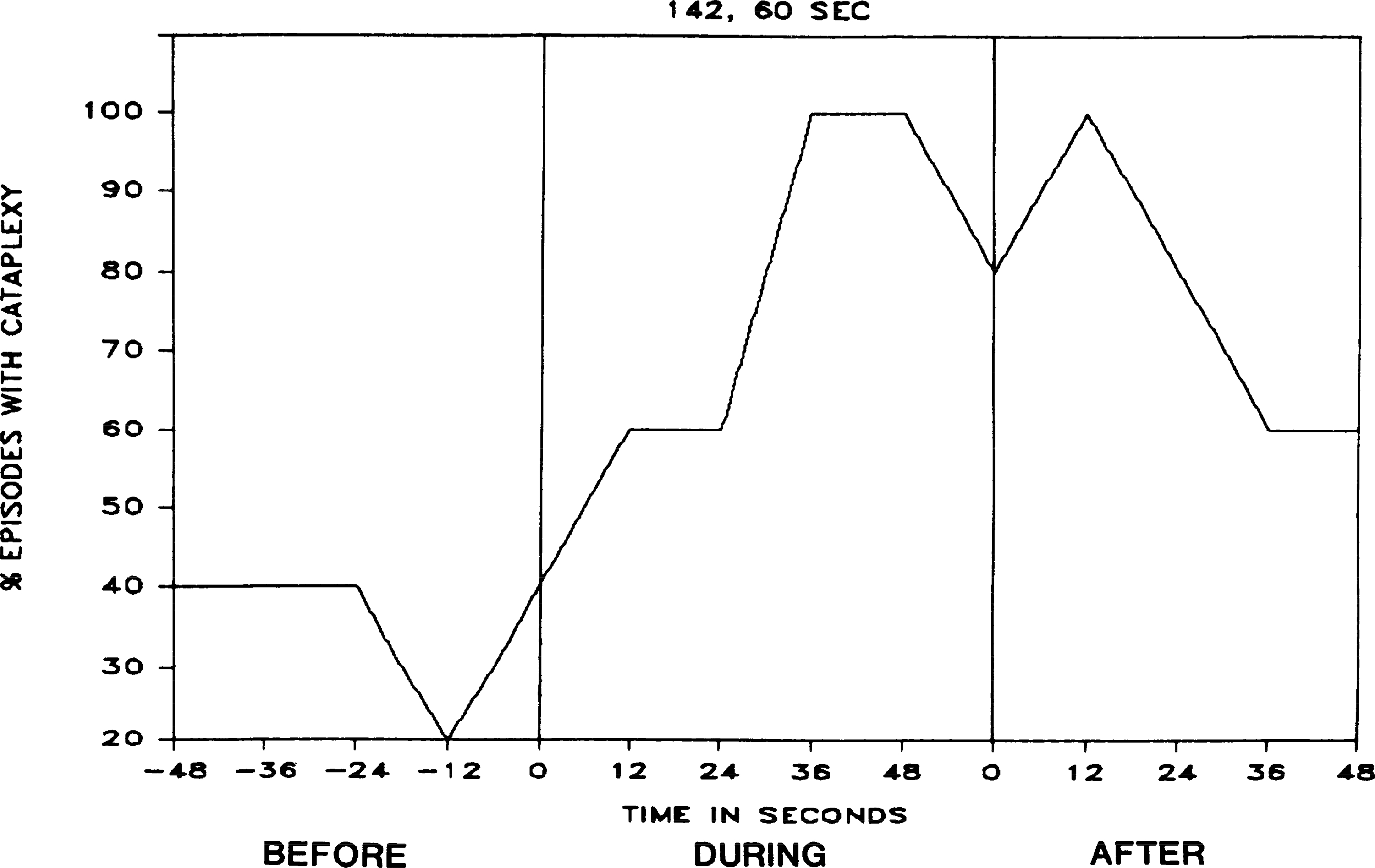
Cataplexy is the abrupt loss of muscle tone experienced by narcoleptics. It is usually precipitated by strong emotions or athletic activity. It has been hypothesized that cardiovascular variables have a role in the triggering of cataplexy. In the present study, we have utilized the narcoleptic canine model to directly investigate changes in heart rate and blood pressure in relation to cataplectic episodes. We found that heart rate increased 18% on average in the 20 s preceding cataplexy onset and then fell during cataplexy. Thus, from a cardiovascular standpoint, cataplexy can be subdivided into two very different periods, the cataplexy onset period with very high and declining heart rate, and the period greater than or equal to 10 s after onset, with greatly reduced heart rate. Heart rate at cataplexy onset was significantly higher than heart rate in rapid-eye-movement (REM) sleep, non-REM sleep, and quiet waking. Blood pressure did not markedly change before the onset of spontaneous cataplexies but decreased significantly during cataplexy. Although blood pressure increases did not precede spontaneous cataplexies, sudden increases in blood pressure, induced pharmacologically or by obstruction of the descending aorta, triggered cataplexy in the most severely affected subjects. A hypothesized role for cataplexy as a homeostatic reflex, triggered by interactions between blood flow, central chemoreceptors, and atonia control mechanisms in the medial medulla, is discussed.
Figures
Similar articles
-
Activity of medial mesopontine units during cataplexy and sleep-waking states in the narcoleptic dog.J Neurosci. 1992 May;12(5):1640-6. doi: 10.1523/JNEUROSCI.12-05-01640.1992. J Neurosci. 1992. PMID: 1578258 Free PMC article.
-
Heart rate and blood pressure changes associated with cataplexy in canine narcolepsy.Sleep. 1986;9(1 Pt 2):216-21. doi: 10.1093/sleep/9.1.216. Sleep. 1986. PMID: 3704445 Free PMC article.
-
Is narcolepsy a REM sleep disorder? Analysis of sleep abnormalities in narcoleptic Dobermans.Neurosci Res. 2000 Dec;38(4):437-46. doi: 10.1016/s0168-0102(00)00195-4. Neurosci Res. 2000. PMID: 11164570
-
The neuronal network responsible for paradoxical sleep and its dysfunctions causing narcolepsy and rapid eye movement (REM) behavior disorder.Sleep Med Rev. 2011 Jun;15(3):153-63. doi: 10.1016/j.smrv.2010.08.002. Epub 2010 Nov 5. Sleep Med Rev. 2011. PMID: 21115377 Review.
-
Physiology of REM sleep, cataplexy, and sleep paralysis.Adv Neurol. 1995;67:245-71. Adv Neurol. 1995. PMID: 8848973 Review.
Cited by
-
A consensus definition of cataplexy in mouse models of narcolepsy.Sleep. 2009 Jan;32(1):111-6. Sleep. 2009. PMID: 19189786 Free PMC article.
-
Narcolepsy: neural mechanisms of sleepiness and cataplexy.J Neurosci. 2012 Sep 5;32(36):12305-11. doi: 10.1523/JNEUROSCI.2630-12.2012. J Neurosci. 2012. PMID: 22956821 Free PMC article. Review. No abstract available.
-
Transfer learning from ECG to PPG for improved sleep staging from wrist-worn wearables.Physiol Meas. 2021 May 13;42(4):10.1088/1361-6579/abf1b0. doi: 10.1088/1361-6579/abf1b0. Physiol Meas. 2021. PMID: 33761477 Free PMC article.
-
Orexin/hypocretin system dysfunction in patients with Takotsubo syndrome: A novel pathophysiological explanation.Front Cardiovasc Med. 2022 Nov 3;9:1016369. doi: 10.3389/fcvm.2022.1016369. eCollection 2022. Front Cardiovasc Med. 2022. PMID: 36407467 Free PMC article.
-
Role of the hypocretin (orexin) receptor 2 (Hcrt-r2) in the regulation of hypocretin level and cataplexy.J Neurosci. 2011 Apr 27;31(17):6305-10. doi: 10.1523/JNEUROSCI.0365-11.2011. J Neurosci. 2011. PMID: 21525270 Free PMC article.
References
-
- Ahmad HR, and Loeschcke HH. Transient and steady state responses of pulmonary ventilation to the medullary extracellular pH after approximately rectangular changes in alveolar Pco2. Pfluegers Arch. 395: 285–292, 1982. - PubMed
-
- Baker TL, and Dement WC. Canine narcolepsy-cataplexy syndrome: evidence for an inherited monoaminergic-cholinergic imbalance. In: Brain Mechanisms of Sleep, edited by Mcginty DJ, Drucker-Colin R, Morrison A, and Parmeggiani PL. New York: Raven, 1985, p. 199–234.
-
- Baust W, Boehmke J, and Blossfeld U. Somato-sympathetic reflexes during natural sleep and wakefulness in unrestrained cats. Exp. Brain Res 12: 361–369, 1971. - PubMed
-
- Birzis L, and Tachibana S. Local cerebral impedance and blood flow during sleep and arousal. Exp. Neurol 9: 269–285, 1964. - PubMed
-
- Colley PS, and Sivarajan M. Regional blood flow in dogs during halothane anesthesia and controlled hypotension produced by nitroprusside or nitroglycerin. Anesth. Analg 63: 503–510, 1984. - PubMed
Publication types
MeSH terms
Substances
Grants and funding
LinkOut - more resources
Full Text Sources