Comparative Study
. 2017 Nov 9;17(1):730.
doi: 10.1186/s12885-017-3737-z.

Detection of circulating miRNAs: comparative analysis of extracellular vesicle-incorporated miRNAs and cell-free miRNAs in whole plasma of prostate cancer patients

Affiliations
Comparative Study

Detection of circulating miRNAs: comparative analysis of extracellular vesicle-incorporated miRNAs and cell-free miRNAs in whole plasma of prostate cancer patients

Edgars Endzeliņš et al. BMC Cancer. .

Abstract

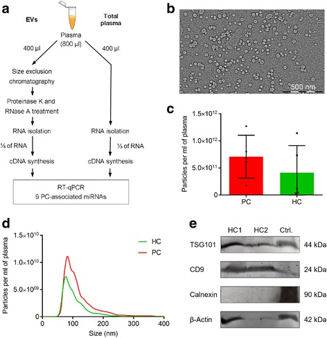
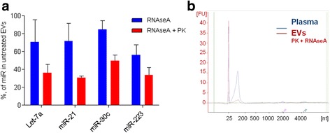
Background: Circulating cell-free miRNAs have emerged as promising minimally-invasive biomarkers for early detection, prognosis and monitoring of cancer. They can exist in the bloodstream incorporated into extracellular vesicles (EVs) and ribonucleoprotein complexes. However, it is still debated if EVs contain biologically meaningful amounts of miRNAs and may provide a better source of miRNA biomarkers than whole plasma. The aim of this study was to systematically compare the diagnostic potential of prostate cancer-associated miRNAs in whole plasma and in plasma EVs.

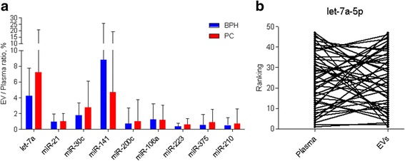
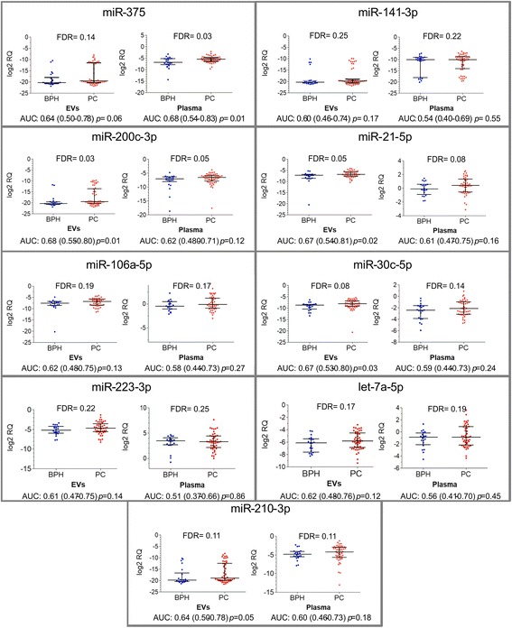
Methods: RNA was isolated from whole plasma and plasma EV samples from a well characterised cohort of 50 patient with prostate cancer (PC) and 22 patients with benign prostatic hyperplasia (BPH). Nine miRNAs known to have a diagnostic potential for PC in cell-free blood were quantified by RT-qPCR and the relative quantities were compared between patients with PC and BPH and between PC patients with Gleason score ≥ 8 and ≤6.

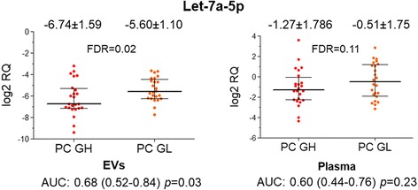
Results: Only a small fraction of the total cell-free miRNA was recovered from the plasma EVs, however the EV-incorporated and whole plasma cell-free miRNA profiles were clearly different. Four of the miRNAs analysed showed a diagnostic potential in our patient cohort. MiR-375 could differentiate between PC and BPH patients when analysed in the whole plasma, while miR-200c-3p and miR-21-5p performed better when analysed in plasma EVs. EV-incorporated but not whole plasma Let-7a-5p level could distinguish PC patients with Gleason score ≥ 8 vs ≤6.

Conclusions: This study demonstrates that for some miRNA biomarkers EVs provide a more consistent source of RNA than whole plasma, while other miRNAs show better diagnostic performance when tested in the whole plasma.

Keywords: Biomarkers; Cell-free miRNAs; Exosomes; Extracellular vesicles; Liquid biopsy; Microvesicles; Prostate cancer.


Conflict of interest statement

Ethics approval and consent to participate

Biobanking procedures were approved by the Committee of Medical Ethics of Latvia (decision No.5, 16.09.2010) and the use of clinical samples for research was approved by the Committee of Biomedical Ethics of Riga East University Hospital (decision No. 7-A/15, 04.06.2015). The blood samples were collected after the patients’ informed written consent was obtained.

Consent for publication

Not applicable.

Competing interests

The authors declare that they have no competing interests.

Publisher’s Note

Springer Nature remains neutral with regard to jurisdictional claims in published maps and institutional affiliations.

Figures

Fig. 1
Fig. 1
Workflow of the study and characterisation of plasma EVs. a Workflow of the study. b Representative transmission electron microscopy image of plasma EVs. c Quantification of EVs isolated from plasma of PC patients and healthy controls (HC) by nanoparticle tracking analysis. d Average size distribution of EVs isolated from plasma of PC patients and healthy controls. e Western blot analysis of EV markers (TSG101, CD9), endoplasmic reticulum protein Calnexin and β-actin in plasma EVs isolated from two healthy individuals and PC-3 cells (as a positive control)
Fig. 2
Fig. 2
Effects of proteinase K and RNase A treatment on the relative quantity of EV-incorporated miRNAs and RNA profiles in whole plasma and EVs. a RT-qPCR analysis of miRNA levels in EVs treated with RNase A alone or with a combination of proteinase K and RNase A relatively to untreated EVs. Bars show the mean percentage in EVs from 3 healthy individuals. b A representative RNA profile from whole plasma and EVs treated with proteinase K and RNase A obtained by Bioanlyzer RNA 6000 Pico chip
Fig. 3
Fig. 3
Relative abundance of EV-incorporated miRNAs. a Ratio between EV-incorporated and total cell-free miRNAs in whole plasma. Bars represent the mean ratios in groups of patients with PC and BPH. b A paired dot plot shows the ranking of PC patients according to Let-7a-5p levels in EVs and whole plasma; lines connect the samples from the same individual
Fig. 4
Fig. 4
Circulating miRNA levels in patients with BPH and PC. Scatter plots show the log2RQ values of each miRNA tested in EVs and in whole plasma. FDR-adjusted p values are show at the top of each graph. Area under the ROC curve (AUC), 95% confidence interval and p value for differentiating between PC and BPH is shown below each graph
Fig. 5
Fig. 5
Circulating Let-7a-5p levels in PC patients with low and high Gleason score. Scatter plots show the log2RQ values of Let-7a-5p tested in EVs and in whole plasma of patients with Gleason score ≥ 8 (PC GH) and Gleason score ≤6 (PC GL). The mean log2RQ values and standard deviation is shown above each scatter plot. Area under the ROC curve (AUC), 95% confidence interval and p value for differentiating between PC patients with high and low Gleason score is shown below each graph

References

    1. Mitchell PS, Parkin RK, Kroh EM, Fritz BR, Wyman SK, Pogosova-Agadjanyan EL, et al. Circulating microRNAs as stable blood-based markers for cancer detection. Proc Natl Acad Sci USA. 2008;105:10513–10518. doi: 10.1073/pnas.0804549105. - DOI - PMC - PubMed
    1. Chen X, Ba Y, Ma L, Cai X, Yin Y, Wang K, et al. Characterization of microRNAs in serum: a novel class of biomarkers for diagnosis of cancer and other diseases. Cell Res. 2008;18:997–1006. doi: 10.1038/cr.2008.282. - DOI - PubMed
    1. Armand-Labit V, Pradines A. Circulating cell-free microRNAs as clinical cancer biomarkers. Biomol Concepts. 2017;8:61–81. doi: 10.1515/bmc-2017-0002. - DOI - PubMed
    1. Siegel RL, Miller KD, Jemal A. Cancer statistics, 2017. CA Cancer J Clin. 2017;67:7–30. doi: 10.3322/caac.21387. - DOI - PubMed
    1. Ferlay J, Soerjomataram I, Dikshit R, Eser S, Mathers C, Rebelo M, et al. Cancer incidence and mortality worldwide: sources, methods and major patterns in GLOBOCAN 2012. Int J Cancer. 2015;136:E359–E386. doi: 10.1002/ijc.29210. - DOI - PubMed

Publication types