

p21-/- mice exhibit enhanced bone regeneration after injury
- PMID: 29121899
- PMCID: PMC5679350
- DOI: 10.1186/s12891-017-1790-z
p21-/- mice exhibit enhanced bone regeneration after injury
Abstract
Background: p21(WAF1/CIP1/SDI1), a cyclin dependent kinase inhibitor has been shown to influence cell proliferation, differentiation and apoptosis; but more recently, p21 has been implicated in tissue repair. Studies on p21(-/-) knockout mice have demonstrated results that vary from complete regeneration and healing of tissue to attenuated healing. There have however been no studies that have evaluated the role of p21 inhibition in bone healing and remodeling.
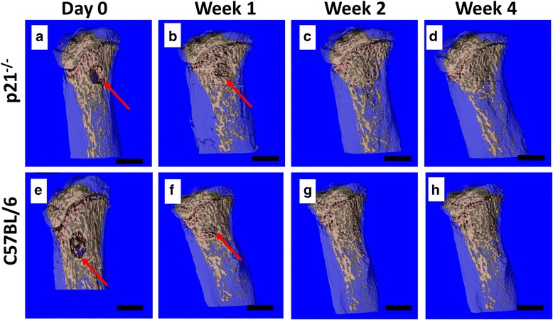
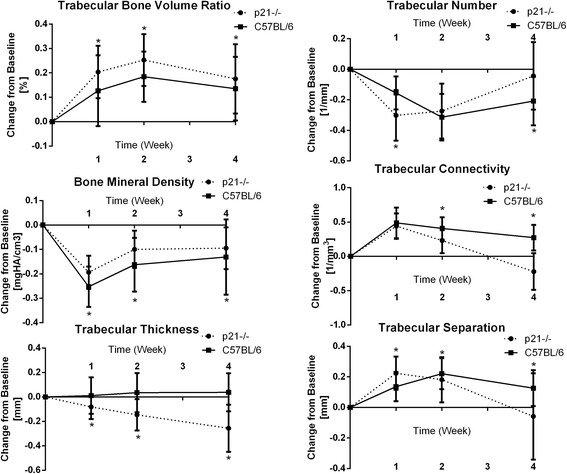
Methods: The current study employs a burr-hole fracture model to investigate bone regeneration subsequent to an injury in a p21-/- mouse model. p21-/- and C57BL/6 mice were subjected to a burr-hole fracture on their proximal tibia, and their bony parameters were measured over 4 weeks via in vivo μCT scanning.
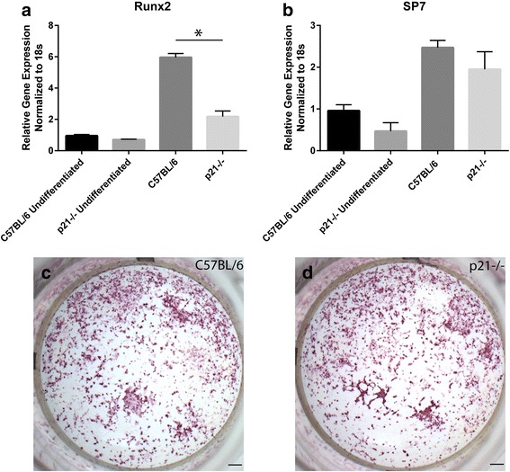
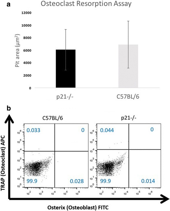
Results: p21-/- mice present with enhanced healing from week 1 through week 4. Differences in bone formation and resorption potential between the two mouse models are assessed via quantitative and functional assays. While the μCT analysis indicates that p21-/- mice have enhanced bone healing capabilities, it appears that the differences observed may not be due to the function of osteoblasts or osteoclasts. Furthermore, no differences were observed in the differentiation of progenitor cells (mesenchymal or monocytic) into osteoblasts or osteoclasts respectively.
Conclusions: Therefore, it remains unknown how p21 is regulating enhanced fracture repair and further studies are required to determine which cell type(s) are responsible for this regenerative phenotype.
Keywords: Bone healing; Mice; Trabecular bone; p21; μCT.
Conflict of interest statement
Ethics approval
All animal procedures are approved by the University of Calgary Animal Care Committee.
Consent for publication
Not applicable.
Competing interests
RK is a member of the Editorial Board of BMC Musculoskeletal Disorders. The other authors declare they have no other competing interest.
Publisher’s Note
Springer Nature remains neutral with regard to jurisdictional claims in published maps and institutional affiliations.
Figures
Similar articles
-
Cell cycle regulators and bone: development and regeneration.Cell Biosci. 2023 Feb 21;13(1):35. doi: 10.1186/s13578-023-00988-7. Cell Biosci. 2023. PMID: 36810262 Free PMC article. Review.
-
Absence of p21(WAF1/CIP1/SDI1) protects against osteopenia and minimizes bone loss after ovariectomy in a mouse model.PLoS One. 2019 Apr 10;14(4):e0215018. doi: 10.1371/journal.pone.0215018. eCollection 2019. PLoS One. 2019. PMID: 30970032 Free PMC article.
-
Absence of E2f1 Negates Pro-osteogenic Impacts of p21 Absence.Calcif Tissue Int. 2024 Jun;114(6):625-637. doi: 10.1007/s00223-024-01210-7. Epub 2024 Apr 21. Calcif Tissue Int. 2024. PMID: 38643416
-
Does Sclerostin Depletion Stimulate Fracture Healing in a Mouse Model?Clin Orthop Relat Res. 2016 May;474(5):1294-302. doi: 10.1007/s11999-015-4640-z. Epub 2015 Nov 25. Clin Orthop Relat Res. 2016. PMID: 26608966 Free PMC article.
-
[A comparative study on effect of different defect diameters on healing in middle 1/3 tibia monolayer cortical bone defect mouse model].Zhongguo Xiu Fu Chong Jian Wai Ke Za Zhi. 2012 Oct;26(10):1218-22. Zhongguo Xiu Fu Chong Jian Wai Ke Za Zhi. 2012. PMID: 23167107 Chinese.
Cited by
-
MiR-1908/EXO1 and MiR-203a/FOS, regulated by scd1, are associated with fracture risk and bone health in postmenopausal diabetic women.Aging (Albany NY). 2020 May 26;12(10):9549-9584. doi: 10.18632/aging.103227. Epub 2020 May 26. Aging (Albany NY). 2020. PMID: 32454462 Free PMC article.
-
Cell cycle regulators and bone: development and regeneration.Cell Biosci. 2023 Feb 21;13(1):35. doi: 10.1186/s13578-023-00988-7. Cell Biosci. 2023. PMID: 36810262 Free PMC article. Review.
-
CircRNA_25487 inhibits bone repair in trauma-induced osteonecrosis of femoral head by sponging miR-134-3p through p21.Regen Ther. 2020 Dec 28;16:23-31. doi: 10.1016/j.reth.2020.12.003. eCollection 2021 Mar. Regen Ther. 2020. PMID: 33426239 Free PMC article.
-
p21-/- Mice Exhibit Spontaneous Articular Cartilage Regeneration Post-Injury.Cartilage. 2021 Dec;13(2_suppl):1608S-1617S. doi: 10.1177/1947603519876348. Epub 2019 Sep 26. Cartilage. 2021. PMID: 31556320 Free PMC article.
-
Integrated computational and in vivo models reveal Key Insights into macrophage behavior during bone healing.PLoS Comput Biol. 2022 May 13;18(5):e1009839. doi: 10.1371/journal.pcbi.1009839. eCollection 2022 May. PLoS Comput Biol. 2022. PMID: 35559958 Free PMC article.
References
MeSH terms
Substances
Grants and funding
LinkOut - more resources
Full Text Sources
Other Literature Sources
Molecular Biology Databases
