. 2017 Nov 9;14(1):219.
doi: 10.1186/s12974-017-0990-7.

Interleukin-1β signaling in fenestrated capillaries is sufficient to trigger sickness responses in mice

Affiliations

Interleukin-1β signaling in fenestrated capillaries is sufficient to trigger sickness responses in mice

J Gabriel Knoll et al. J Neuroinflammation. .

Abstract

Background: The physiological and behavioral symptoms of sickness, including fever, anorexia, behavioral depression, and weight loss can be both beneficial and detrimental. These sickness responses are triggered by pro-inflammatory cytokines acting on cells within the brain. Previous research demonstrates that the febrile response to peripheral insults depends upon prostaglandin production by vascular endothelial cells, but the mechanisms and specific cell type(s) responsible for other sickness responses remain unknown. The purpose of the present study was to identify which cells within the brain are required for sickness responses triggered by central nervous system inflammation.

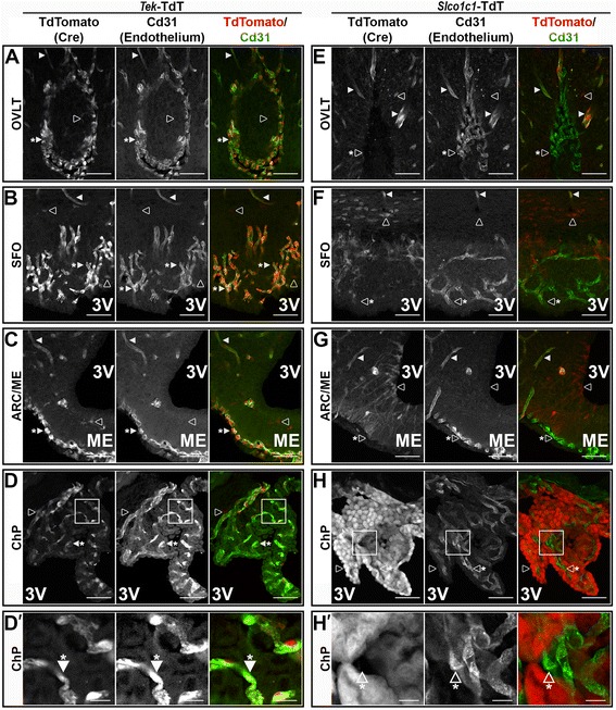
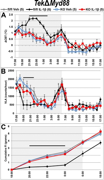
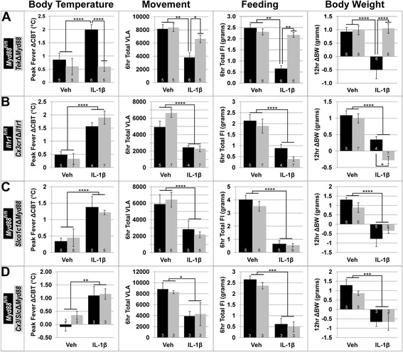
Methods: Intracerebroventricular (ICV) administration of 10 ng of the potent pro-inflammatory cytokine interleukin-1β (IL-1β) was used as an experimental model of central nervous system cytokine production. We examined which cells respond to IL-1β in vivo via fluorescent immunohistochemistry. Using multiple transgenic mouse lines expressing Cre recombinase under the control of cell-specific promoters, we eliminated IL-1β signaling from different populations of cells. Food consumption, body weight, movement, and temperature were recorded in adult male mice and analyzed by two-factor ANOVA to determine where IL-1β signaling is essential for sickness responses.

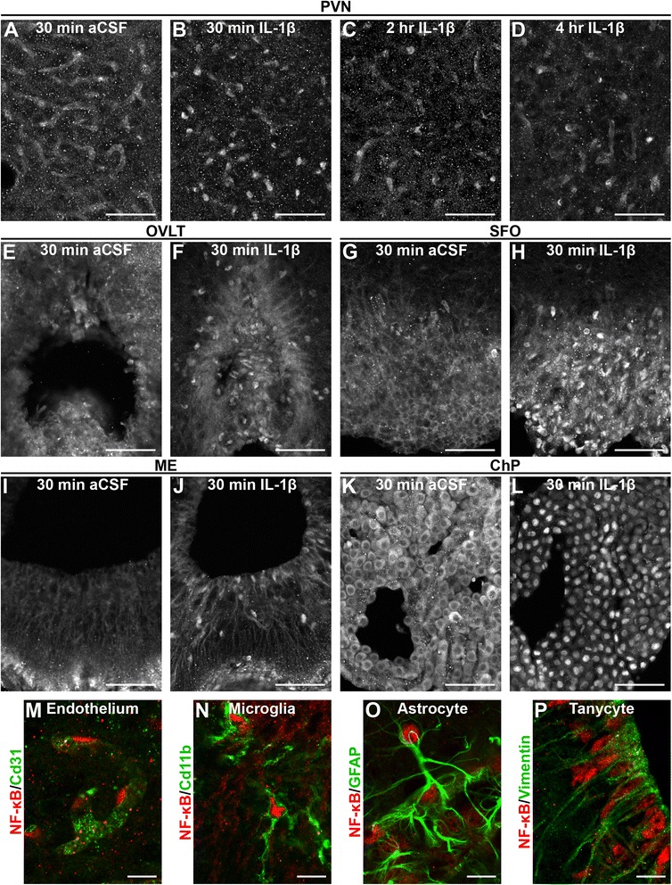
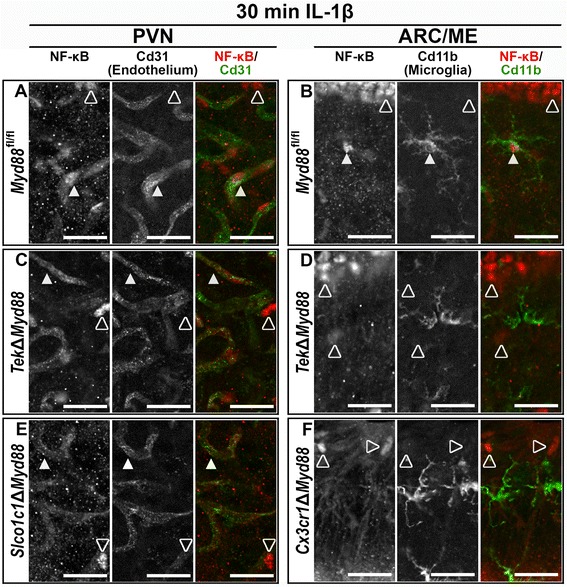
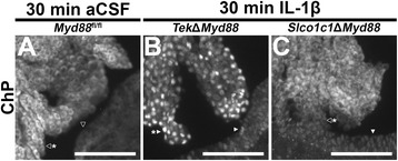
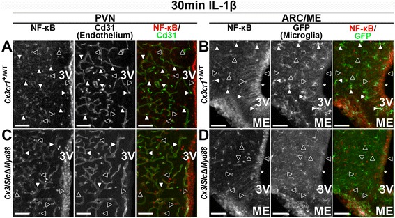
Results: Endothelial cells, microglia, ependymal cells, and astrocytes exhibit nuclear translocation of NF-κB (nuclear factor kappa-light-chain-enhancer of activated B cells) in response to IL-1β. Interfering with IL-1β signaling in microglia, endothelial cells within the parenchyma of the brain, or both did not affect sickness responses. Only mice that lacked IL-1β signaling in all endothelium including fenestrated capillaries lacked sickness responses.

Conclusions: These experiments show that IL-1β-induced sickness responses depend on intact IL-1β signaling in blood vessels and suggest that fenestrated capillaries act as a critical signaling relay between the immune and nervous systems.

Trial registration: Not applicable.

Keywords: Cytokine; Endothelial cells; Fenestrated capillaries; Hypothalamus; Inflammation; Microglia; Sickness behavior.


Conflict of interest statement

Ethics approval

All animal care, handling, and experimentation were conducted in accordance with the OHSU Institutional Animal Care and Use Committee (IACUC) guidelines.

Consent for publication

Not applicable.

Competing interests

The authors declare that they have no competing interests.

Publisher’s Note

Springer Nature remains neutral with regard to jurisdictional claims in published maps and institutional affiliations.

Figures

Fig. 1
Fig. 1
Cd31 immunoreactivity (IR) shows brain vascular heterogeneity. Increased vascular density demonstrated by Cd31 IR in representative epifluorescent images of the organum vasculosum lamina terminalis (OVLT, a), subfornical organ (SFO, b), choroid plexus (ChP, c), paraventricular nucleus (PVN, d), and arcuate nucleus/median eminence (ARC/ME, e). 3V third ventricle. Scale bars = 500 μm
Fig. 2
Fig. 2
Intracerebroventricular (ICV) administration of IL-1β induces NF-κB nuclear localization in a time- and region-dependent manner. The effect of ICV IL-1β is both rapid and transient as demonstrated by NF-κB immunoreactivity (IR) in representative confocal images of the paraventricular nucleus (PVN). IR in vehicle (aCSF, n = 6)-treated animals showed cytoplasmic labeling of blood vessels (a). IL-1β treatment caused NF-κB nuclear localization that peaked by 30 min (b, n = 8), persisted for at least 2 h (c, n = 4) and returned to near baseline, with cytoplasmic vascular patterns reappearing, by 4 h after treatment (d, n = 4). This effect was most prominent in and around the PVN, organum vasculosum lamina terminalis (OVLT; e, f), subfornical organ (SFO; g, h), arcuate nucleus/median eminence (ARC/ME; i, j), and choroid plexus (ChP; k, l). High-magnification confocal images of co-labeling with cell-specific markers shows that vascular endothelial cells (m), microglia (n), astrocytes (o), and tanycytes (p) directly respond to IL-1β. Scale bars: al = 50 μm, mp = 10 μm
Fig. 3
Fig. 3
Recombinase reporter shows strain-specific Cre expression. Representative confocal images demonstrate that Tek-Cre is expressed in Cd31+ vascular endothelial cells (a) and Iba1+ microglia (b, n = 4). Cx3cr1-CreERT2 is only expressed in microglia in both the PVN and ME (c, d, n = 6). Slco1c1-CreERT2 is expressed in blood vessels (e) but not microglia (f, n = 4). TdTomato+ cells with long processes in f are β1 tanycytes. Scale bars = 25 μm
Fig. 4
Fig. 4
Tek-Cre, but not Slco1c1-CreERT2, is expressed in fenestrated capillaries of circumventricular organs. Representative confocal images of co-localization of TdTomato (TdT, red) and Cd31 (green) in Tek-TdT animals (ac, n = 4) confirms co-expression in parenchymal endothelium (filled arrowheads) and fenestrated capillaries (filled arrowheads with asterisk) in the organum vasculosum lamina terminalis (OVLT, a), subfornical organ (SFO, b), arcuate nucleus/median eminence (ARC/ME, c), and choroid plexus (ChP, d). Reporter expression is also seen in microglia in each of these regions (open arrowheads in ad). Co-labeling of TdT (red) and Cd31(green) in Slco1c1-TdT animals (eh, n = 4) shows Cre activity in all parenchymal endothelium (filled arrowheads), but not fenestrated capillaries (open arrowheads with asterisk). Reporter expression is also seen in other cells that do not express Cd31, particularly the cuboidal cells of the choroid plexus (open arrowheads in eh). Higher magnification of the boxed regions in d and h (d′ and h′) demonstrate that ChP fenestrated capillaries express TdT in Tek-Cre but not Slco1c1-CreERT2 animals (filled and open arrowheads with asterisk, respectively). Scale bars: ah 50 μm; d′ and h′ 10 μm
Fig. 5
Fig. 5
NF-κB immunoreactivity (IR) confirms strain-specific IL-1β signaling disruption. Representative confocal images show that in control animals (Myd88 fl/fl), IL-1β causes nuclear localization of NF-κB (red) in endothelial cells (Cd31+, green; filled arrowheads in a and microglia (Cd11b+, green; filled arrowheads in b, n = 3). Nuclear IR was also present in cells that did not express co-labeled proteins (open arrowheads in af). TekΔMyd88 disrupts IL-1β signaling in endothelium and microglia as NF-κB remains cytoplasmic in blood vessels (filled arrows in c) and nuclear IR was absent from microglia (d, n = 3). The vascular pattern of cytoplasmic NF-κB IR shows that Slco1c1ΔMyd88 disrupts signaling in parenchymal endothelium (filled arrowheads in e, n = 3). The absence of nuclear NF-κB in microglia of Cx3cr1ΔMyd88 demonstrates microglia signaling disruption (f, n = 3). Scale bars = 25 μm
Fig. 6
Fig. 6
NF-κB immunoreactivity (IR) in the choroid plexus confirms promotor-specific genetic recombination. Representative epifluorescent images of the choroid plexus (ChP) and neighboring ependyma 30 min after treatment confirms genetic recombination of Myd88 in a pattern consistent with reporter crosses (see Fig. 4a, h). In vehicle-treated (artificial cerebrospinal fluid, aCSF) control animals (Myd88 fl/fl, n = 3; a), clear nuclear voids of NF-κB IR are present in the cuboidal cells of the ChP (open arrowhead with asterisk) and only diffuse, cytoplasmic labeling, without concentrated nuclear labeling, is seen in the ependyma (open arrowhead). As with control animals (see Fig. 2k–l and Additional file 3: Figure S3A, B), nuclear NF-κB is evident in both cuboidal (arrowhead with asterisk in b) and ependymal cells (arrowhead in b) in response to 10 ng intracerebroventricular IL-1β treatment of TekΔMyd88 (n = 3), which do not express Cre in either cell type. In contrast, while nuclear NF-κB is present in the ependymal cells (arrowhead in c) of IL-1β-treated Slco1c1ΔMyd88 animals (n = 3), NF-κB IR remains cytoplasmic in ChP cuboidal cells where Slco1c1-Cre is expressed (arrowhead with asterisk in c; compare with Fig. 4h). Scale bars = 100 μm
Fig. 7
Fig. 7
Combined Cx3/Slc-CreERT2 eliminates IL-1β signaling in parenchymal endothelium and microglia. Representative epifluorescent images of the paraventricular nucleus (PVN, a) and arcuate nucleus/median eminence region (ARC/ME, b) showing IL-1β-induced nuclear NF-κB immunoreactivity (IR) in parenchymal endothelium (co-expression with Cd31 indicated by filled arrowheads in a) and GFP+ microglia (filled arrowheads in b) in Cx3cr1-CreERT2+ animals that do not have floxed Myd88 (Cx3cr1 +/WT, n = 5). Nuclear NF-κB IR is also found in cells that do not express either marker (open arrowheads in a and b), including ependymal cells lining the third ventricle (3V) and β1-tanycytes (asterisk in b). Representative images from an IL-1β-treated combined Cre animal (Cx3/SlcΔMyd88, n = 3) showing cytoplasmic NF-κB IR in PVN parenchymal endothelium (filled arrowheads in c) and an absence of nuclear NF-κB IR in GFP+ microglia (d) demonstrating that these cells do not respond to ICV IL-1β in combined Cre animals. Similar to control animals, nuclear NF-κB IR is found in cells that do not express either marker (open arrowheads in c and d), but with an apparent decrease in β1-tanycytes (asterisk in d) where Slco1c1-CreERT2 is expressed (see Fig. 4g). Scale bars = 50 μm
Fig. 8
Fig. 8
TekΔMyd88-mediated disruption of IL-1β signaling in all endothelium and microglia eliminates sickness response. Twenty-four-hour profiles of telemetric and feeding data shows the stereotypical IL-1β-induced elevation in core body temperature (ΔCBT, a) and decrease in voluntary locomotor activity (VLA, b) and food intake (FI, c) in Myd88 fl/fl (fl/fl) but not TekΔMyd88 (KO) mice. IL-1β-treated control ΔCBT, VLA and FI were significantly different from vehicle (Veh)-treated values for several hours following treatment (p < 0.05 for times below black bar above traces in ac). IL-1β-treated KO animals were not different from Veh-treated animals at any time. Gray boxes show dark phase, when mice are most active. All values shown are mean ± SEM for group sizes listed in the legend above a
Fig. 9
Fig. 9
IL-1β-induced sickness responses are only eliminated when signaling is disrupted in all endothelium and microglia. TekΔMyd88-mediated disruption of IL-1β signaling in all endothelium and microglia eliminates the increase in core body temperature (ΔCBT) and decrease in voluntary locomotor activity (VLA), food intake (FI), and body weight (ΔBW) associated with IL-1β treatment of Myd88 fl/fl mice (a). Targeted disruption of IL-1β signaling in microglia alone (Cx3cr1ΔIl1r1, b), parenchymal endothelium (Slco1c1ΔMyd88, c) or both parenchymal endothelium and microglia (Cx3/SlcΔMyd88, d) was insufficient to alter the sickness response. IL-1β treatment is the only factor that influences ΔCBT, VLA, FI, and ΔBW for all genotypes except TekΔMyd88. All values shown are mean ± SEM for group sizes listed in graph bars. *p < 0.05; **p < 0.01; ***p < 0.001; ****p < 0.0001

Similar articles

Cited by

References

    1. Dantzer R, Kelley KW. Twenty years of research on cytokine-induced sickness behavior. Brain Behav Immun. 2007;21(2):153–60. Epub 2006/11/08. doi: 10.1016/j.bbi.2006.09.006. PubMed PMID: 17088043; PubMed Central PMCID: PMC1850954 - PMC - PubMed
    1. Sohn JW, Elmquist JK, Williams KW. Neuronal circuits that regulate feeding behavior and metabolism. Trends Neurosci. 2013;36(9):504–512. doi: 10.1016/j.tins.2013.05.003. - DOI - PMC - PubMed
    1. Morrison SF. Central control of body temperature. F1000Research. 2016;5. Epub 2016/05/31. doi:10.12688/f1000research.7958.1. PubMed PMID: 27239289; PubMed Central PMCID: PMC4870994. - PMC - PubMed
    1. Holub M, Lawrence DA, Andersen N, Davidova A, Beran O, Maresova V, Chalupa P. Cytokines and chemokines as biomarkers of community-acquired bacterial infection. Mediat Inflamm. 2013;2013:190145. doi: 10.1155/2013/190145. - DOI - PMC - PubMed
    1. Currie HN, Loos MS, Vrana JA, Dragan K, Boyd JW. Spatial cytokine distribution following traumatic injury. Cytokine. 2014;66(2):112–118. doi: 10.1016/j.cyto.2014.01.001. - DOI - PubMed

MeSH terms

Substances

LinkOut - more resources