

Non-invasive quantification of age-related changes in the vertebral endplate in rats using in vivo DCE-MRI
- PMID: 29121960
- PMCID: PMC5680764
- DOI: 10.1186/s13018-017-0669-x
Non-invasive quantification of age-related changes in the vertebral endplate in rats using in vivo DCE-MRI
Abstract
Background: Small animal models that can mimic degenerative disc disease (DDD) are commonly used to examine DDD progression. However, assessments such as histological studies and macroscopic measurements do not allow for longitudinal studies because they can only be completed after the animal is sacrificed. Dynamic contrast-enhanced MRI (DCE-MRI) may provide a reliable, non-invasive in vivo method for detecting the progression.
Methods: The present study investigated the progression of changes in lumbar discs and the effect of endplate conditions on diffusion into the lumbar discs of aging sand rats after intravenous administration of gadolinium-containing contrast medium through the tail vein. Contrast enhancement was measured in the lumbar intervertebral discs on each image. The results were compared with those from conventional histological characterizations.
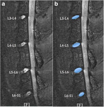
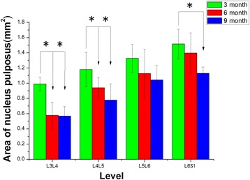
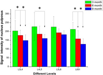
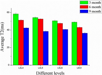
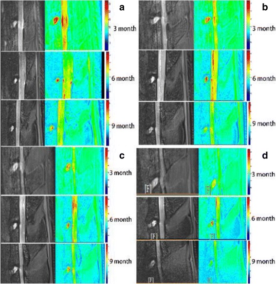
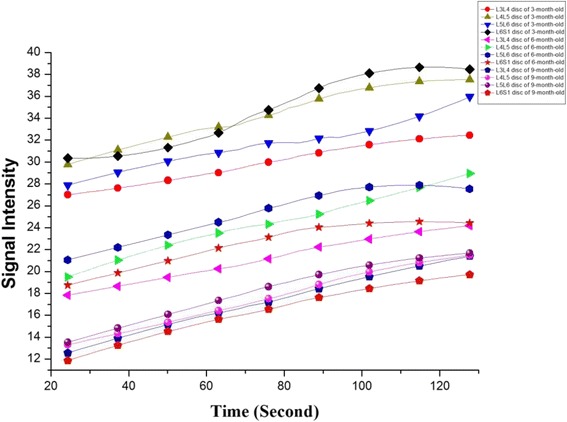
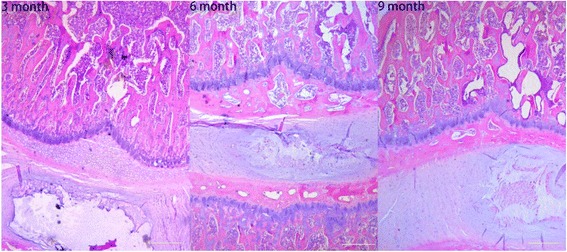
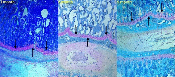
Results: T2-weighted images revealed that with aging, the shape of L3-L4, L4-L5, L5-L6, and L6-S1 nucleus pulposus (NP) became irregular, while the mean areas, signal intensities, and T2 values of the NP were significantly decreased. Each of the observed disc changes demonstrated a progressive increase in phase during 2-min scout scans. Post-contrast MRI showed impaired endplate nutritional diffusion to the disc with aging, enhancement was significantly greater in young animals than in old animals. Endplate calcification or sclerosis was histologically confirmed; histologic score was correlated with the age. We found the histological score of the endplate negatively corresponded to the DCE-MRI results.
Conclusions: DCE-MRI studies offer a non-invasive in vivo method for investigating the progress of diffusion into the discs and the functional conditions of the endplate. We conclude that quantitative DCE-MRI can identify the severity of disc degeneration and efficiently reflect the progression of vertebral endplate changes in the aging sand rat lumbar spine via the NP contrast enhancement patterns.
Keywords: Degenerative disc disease; Dynamic contrast-enhanced magnetic resonance imaging; Intervertebral disc endplate; Lumbar spine.
Conflict of interest statement
Ethics approval
All animal experiments were approved by the Animal Ethical Committee and Neurosurgical Institute of Beijing, The Capital Medical University.
Consent for publication
Not applicable.
Competing interests
The authors declare that they have no competing interests.
Publisher’s Note
Springer Nature remains neutral with regard to jurisdictional claims in published maps and institutional affiliations.
Figures
References
MeSH terms
Substances
Grants and funding
LinkOut - more resources
Full Text Sources
Other Literature Sources
Medical
Research Materials
Miscellaneous
