

Radioprotective agents to prevent cellular damage due to ionizing radiation
- PMID: 29121966
- PMCID: PMC5680756
- DOI: 10.1186/s12967-017-1338-x
Radioprotective agents to prevent cellular damage due to ionizing radiation
Abstract
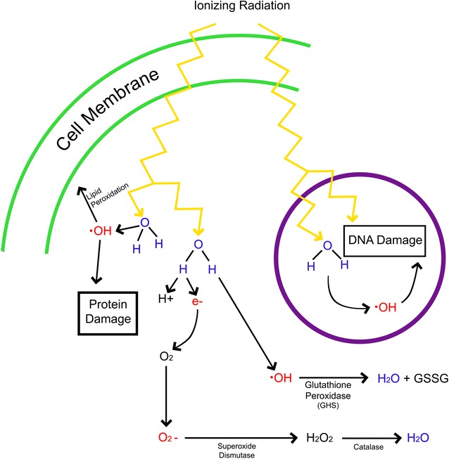
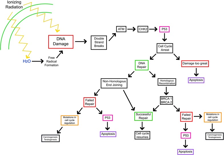
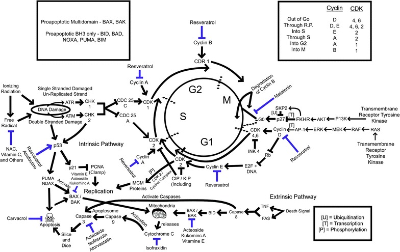
Medical imaging has become a central component of patient care to ensure early and accurate diagnosis. Unfortunately, many imaging modalities use ionizing radiation to generate images. Ionizing radiation even in low doses can cause direct DNA damage and generate reactive oxygen species and free radicals, leading to DNA, protein, and lipid membrane damage. This cell damage can lead to apoptosis, necrosis, teratogenesis, or carcinogenesis. As many as 2% of cancers (and an associated 15,000 deaths annually) can be linked to computed tomography exposure alone. Radioprotective agents have been investigated using various models including cells, animals, and recently humans. The data suggest that radioprotective agents working through a variety of mechanisms have the potential to decrease free radical damage produced by ionizing radiation. Radioprotective agents may be useful as an adjunct to medical imaging to reduced patient morbidity and mortality due to ionizing radiation exposure. Some radioprotective agents can be found in high quantities in antioxidant rich foods, suggesting that a specific diet recommendation could be beneficial in radioprotection.
Keywords: Antioxidant; Computed tomography; Ionizing radiation; Medical imaging; Mitigators; Radioprotectant.
Figures
Similar articles
-
Review of compounds that exhibit radioprotective and/or mitigatory effects after application of diagnostic or therapeutic ionizing radiation.Int J Radiat Biol. 2023;99(4):594-603. doi: 10.1080/09553002.2022.2110308. Epub 2022 Aug 17. Int J Radiat Biol. 2023. PMID: 35930681 Review.
-
Radioprotective countermeasures for radiation injury (Review).Mol Med Rep. 2023 Mar;27(3):66. doi: 10.3892/mmr.2023.12953. Epub 2023 Feb 17. Mol Med Rep. 2023. PMID: 36799170 Free PMC article. Review.
-
Potential utility of peptides against damage induced by ionizing radiation.Future Oncol. 2021 Apr;17(10):1219-1235. doi: 10.2217/fon-2020-0577. Epub 2021 Feb 17. Future Oncol. 2021. PMID: 33593084 Review.
-
Radioprotective agents for radiation therapy: future trends.Future Oncol. 2014 Dec;10(15):2345-57. doi: 10.2217/fon.14.175. Future Oncol. 2014. PMID: 25525844 Review.
-
Molecular Hydrogen as a Potential Clinically Applicable Radioprotective Agent.Int J Mol Sci. 2021 Apr 27;22(9):4566. doi: 10.3390/ijms22094566. Int J Mol Sci. 2021. PMID: 33925430 Free PMC article. Review.
Cited by
-
Comparative Analysis of Cystamine and Cysteamine as Radioprotectors and Antioxidants: Insights from Monte Carlo Chemical Modeling under High Linear Energy Transfer Radiation and High Dose Rates.Int J Mol Sci. 2024 Sep 29;25(19):10490. doi: 10.3390/ijms251910490. Int J Mol Sci. 2024. PMID: 39408820 Free PMC article.
-
Sex Differences in X-ray-Induced Endothelial Damage: Effect of Taurine and N-Acetylcysteine.Antioxidants (Basel). 2022 Dec 29;12(1):77. doi: 10.3390/antiox12010077. Antioxidants (Basel). 2022. PMID: 36670939 Free PMC article.
-
Parathyroid Hormone 1 Receptor Signaling in Dental Mesenchymal Stem Cells: Basic and Clinical Implications.Front Cell Dev Biol. 2021 Oct 25;9:654715. doi: 10.3389/fcell.2021.654715. eCollection 2021. Front Cell Dev Biol. 2021. PMID: 34760881 Free PMC article. Review.
-
MicroRNA: a novel implication for damage and protection against ionizing radiation.Environ Sci Pollut Res Int. 2021 Apr;28(13):15584-15596. doi: 10.1007/s11356-021-12509-5. Epub 2021 Feb 3. Environ Sci Pollut Res Int. 2021. PMID: 33533004 Free PMC article. Review.
-
Genoprotective activities of plant natural substances in cancer and chemopreventive strategies in the context of 3P medicine.EPMA J. 2020 May 29;11(2):261-287. doi: 10.1007/s13167-020-00210-5. eCollection 2020 Jun. EPMA J. 2020. PMID: 32547652 Free PMC article. Review.
References
-
- Xiao L, Tsutsui T, Miwa N. The lipophilic vitamin C derivative, 6-o-palmitoylascorbate, protects human lymphocytes, preferentially over ascorbate, against X-ray-induced DNA damage, lipid peroxidation, and protein carbonylation. Mol Cell Biochem. 2014;394(1–2):247–259. doi: 10.1007/s11010-014-2101-8. - DOI - PubMed
Publication types
MeSH terms
Substances
Grants and funding
LinkOut - more resources
Full Text Sources
Other Literature Sources
Research Materials
