. 2017 Nov 9;14(1):217.
doi: 10.1186/s12974-017-0987-2.

Endothelial α6β4 integrin protects during experimental autoimmune encephalomyelitis-induced neuroinflammation by maintaining vascular integrity and tight junction protein expression

Affiliations

Endothelial α6β4 integrin protects during experimental autoimmune encephalomyelitis-induced neuroinflammation by maintaining vascular integrity and tight junction protein expression

Jennifer V Welser et al. J Neuroinflammation. .

Abstract

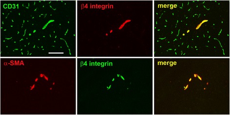
Background: Extracellular matrix (ECM) proteins play critical functions regulating vascular formation and function. Laminin is a major component of the vascular basal lamina, and transgenic mice deficient in astrocyte or pericyte laminin show defective blood-brain barrier (BBB) integrity, indicating an important instructive role for laminin in cerebral blood vessels. As previous work shows that in the normal brain, vascular expression of the laminin receptor α6β4 integrin is predominantly restricted to arterioles, but induced on all vessels during neuroinflammation, it is important to define the role of this integrin in the maintenance of BBB integrity.

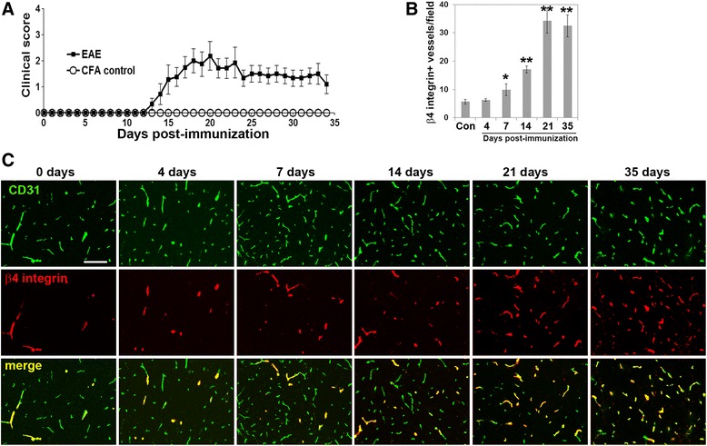
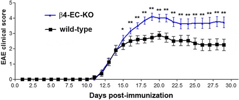
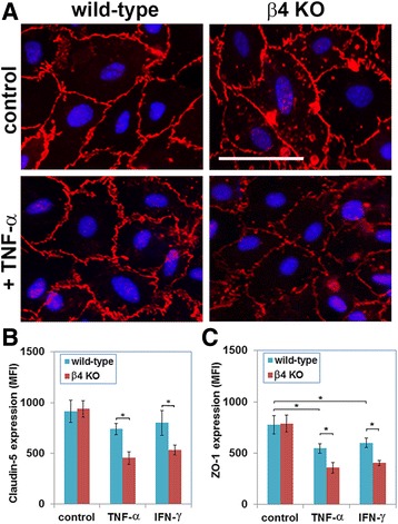
Methods: α6β4 integrin expression was analyzed using dual immunofluorescence (dual-IF) of brain sections taken from the mouse model of multiple sclerosis, experimental autoimmune encephalomyelitis (EAE). To investigate the role of endothelial α6β4 integrin, transgenic mice lacking β4 integrin in endothelial cells (β4-EC-KO) and wild-type (WT) littermates were subject to EAE, and clinical score and various neuropathological parameters were examined by immunofluorescence. In addition, β4 integrin null brain endothelial cells (BECs) were examined in culture for expression of tight junction proteins using immunocytochemistry and flow cytometry.

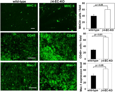
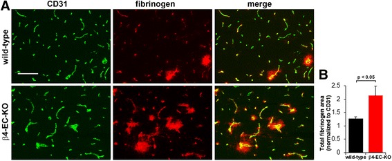
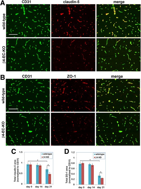
Results: Cerebrovascular expression of β4 integrin was markedly upregulated during EAE progression, such that by the acute stage of EAE (day 21), the vast majority of blood vessels expressed β4 integrin. In the EAE model, while the β4-EC-KO mice showed the same time of disease onset as the WT littermates, they developed significantly worse clinical disease over time, resulting in increased clinical score at the peak of disease and maintained elevated thereafter. Consistent with this, the β4-EC-KO mice showed enhanced levels of leukocyte infiltration and BBB breakdown and also displayed increased loss of the endothelial tight junction proteins claudin-5 and ZO-1. Under pro-inflammatory conditions, primary cultures of β4KO BECs also showed increased loss of claudin-5 and ZO-1 expression.

Conclusions: Taken together, our data suggest that α6β4 integrin upregulation is an inducible protective mechanism that stabilizes the BBB during neuroinflammatory conditions.

Keywords: Blood-brain barrier; Endothelial; Extracellular matrix; Integrin; Laminin; Vascular.


Conflict of interest statement

Ethics approval

The studies described have been reviewed and approved by The Scripps Research Institute Institutional Animal Care and Use Committee.

Consent for publication

Not applicable

Competing interests

The authors declare that they have no competing interests.

Publisher’s Note

Springer Nature remains neutral with regard to jurisdictional claims in published maps and institutional affiliations.

Figures

Fig. 1
Fig. 1
Characterization of β4 integrin expression on blood vessels in the normal brain. Top panel shows dual-IF on frozen sections of the medulla oblongata from adult mice using antibodies specific for the endothelial marker CD31 (AlexaFluor-488, green) and β4 integrin (Cy3, red). Lower panel shows dual-IF using antibodies specific for smooth muscle cell marker α-SMA (Cy3, red) and β4 integrin (AlexaFluor-488, green). Scale bar = 100 μm. Note that β4 integrin was expressed by only a fraction of CD31-positive vessels, but co-localized strongly with α-SMA
Fig. 2
Fig. 2
Vascular β4 integrin expression is strongly upregulated during the development of EAE. a Time course of increasing EAE severity (clinical score) with time post-immunization. b Quantification of the number of blood vessels expressing β4 integrin in frozen sections of the medulla oblongata during EAE progression. Note that significant upregulation of endothelial β4 integrin was detected 7 days post-immunization and peaked after 21 days. *p < 0.05, **p < 0.01. c Frozen sections of the medulla oblongata at different time points of EAE progression were dual-stained using antibodies specific for the endothelial marker CD31 (AlexaFluor-488, green) and β4 integrin (Cy3, red). Scale bar = 100 μm. Note that in the disease-free brain (0 days), β4 integrin was expressed by only a fraction of CD31-positive vessels, but the number of vessels expressing β4 integrin gradually increased with EAE progression, such that by the acute stage of EAE (day 21), the vast majority of blood vessels expressed β4 integrin, and this expression was maintained high through to the chronic stage (day 35)
Fig. 3
Fig. 3
The impact of genetically deleting endothelial β4 integrin on clinical severity in EAE. The progression of EAE in the β4-EC-KO and WT littermate control mice was evaluated by measuring clinical score on daily intervals. All points represent the mean ± SD (n = 16 mice per strain). Note that while the β4-EC-KO mice showed the same time of disease onset as the WT littermates, over time, they developed significantly worse clinical disease, resulting in increased mean clinical score for the duration of the experiment. This result was confirmed in three separate experiments. *p < 0.05, **p < 0.01
Fig. 4
Fig. 4
The impact of genetically deleting endothelial β4 integrin on neuroinflammation in EAE. Frozen brain sections taken from the β4-EC-KO and WT littermate control mice at the acute stage of EAE (day 21) were stained using antibodies specific for the inflammatory leukocyte markers MHC II, CD45, and Mac-1. Data points represent the mean ± SEM of events observed in the medulla oblongata (n = 4 mice). Scale bar = 100 μm. Note that quantification of all three markers revealed that β4-EC-KO brains contained increased numbers of MHC II+ and CD45+ inflammatory leukocytes, as well as increased expression level of the microglial/macrophage marker Mac-1
Fig. 5
Fig. 5
Evaluating the impact of genetically deleting endothelial β4 integrin on BBB integrity in EAE. a Frozen sections of the medulla oblongata taken from the β4-EC-KO and WT littermate control mice at the acute stage of EAE (day 21) were dual-stained using antibodies specific for the endothelial marker CD31 (AlexaFluor-488, green) and fibrinogen (Cy3, red). Scale bar = 100 μm. b Quantification of fibrinogen leakage in the β4-EC-KO vs. WT littermate mice. Data points represent the mean ± SEM of events observed in the medulla oblongata (n = 4 mice). Scale bar = 100 μm. Note that that in most blood vessels, fibrinogen staining was localized to within the lumen of blood vessels. However, in some blood vessels, fibrinogen staining extended beyond the vascular margin, creating a fuzzy leakage pattern, indicative of loss of vascular integrity. Also note that in the β4-EC-KO mice, fibrinogen leakage was noticeably worse than the WT controls, both in the number and extent of leaky vessels, and this was confirmed by quantification
Fig. 6
Fig. 6
Evaluating the impact of genetic deletion of endothelial β4 integrin on endothelial tight junction protein expression in EAE. a, b Frozen sections of the medulla oblongata taken from the β4-EC-KO and WT littermate control mice at the acute stage of EAE (day 21) were dual-stained using antibodies specific for the endothelial marker CD31 (AlexaFluor-488, green) and claudin-5 (Cy3, red) or CD31 (AlexaFluor-488, green) and ZO-1 (Cy3, red) in A and B, respectively. Scale bar = 100 μm. c, d Quantification of endothelial expression of claudin-5 (c) and ZO-1 (d) in the β4-EC-KO vs. WT littermate mice. Data points represent the mean ± SEM of events observed in the medulla oblongata (n = 4 mice). Note that in the WT mice, endothelial levels of claudin-5 and ZO-1 were markedly reduced in the acute phase of EAE (day 21), and at this phase of disease, expression levels in the β4-EC-KO mice were significantly reduced compared to their WT littermates
Fig. 7
Fig. 7
Evaluating endothelial tight junction protein expression in primary brain endothelial cells (BECs). a BECs from the β4-EC-KO and WT littermate control mice were cultured on laminin in the presence or absence of TNF-α, and claudin-5 expression was examined by IF. b, c Quantification of claudin-5 (b) or ZO-1 (c) expression by β4 integrin null and WT BECs under pro-inflammatory conditions (treatment with TNF-α or IFN-γ). All points represent the mean ± SEM of the mean fluorescent intensity (MFI) of three separate experiments. Note that while ZO-1 expression in WT BECs was significantly reduced by TNF-α and IFN-γ, expression of claudin-5 was not significantly affected by either of these cytokines. Furthermore, compared to WT BECs, while β4 integrin KO cells expressed equivalent levels of claudin-5 and ZO-1 under basal conditions, after exposure to TNF-α or IFN−γ, β4 integrin KO BECs expressed significantly lower levels of claudin-5 and ZO-1. *p < 0.05

Similar articles

Cited by

References

    1. Ffrench-Constant C. Pathogenesis of multiple sclerosis. Lancet. 1994;343:271–275. doi: 10.1016/S0140-6736(94)91118-5. - DOI - PubMed
    1. Lassmann H. Multiple sclerosis pathology. In: Compston A, editor. McAlpine’s multiple sclerosis Churchill Livingstone. 3. 1998. pp. 323–358.
    1. Gay D, Esiri M. Blood–brain barrier damage in acute multiple sclerosis plaques. Brain. 1991;114:557–572. doi: 10.1093/brain/114.1.557. - DOI - PubMed
    1. Kirk J, Plumb J, Mirakhur M, McQuaid S. Tight junction abnormality in multiple sclerosis white matter affects all calibres of vessel and is associated with blood–brain barrier leakage and active demyelination. J Pathol. 2003;201:319–327. doi: 10.1002/path.1434. - DOI - PubMed
    1. Roscoe WA, Welsh ME, Carter DE, Karlik SJ. VEGF and angiogenesis in acute and chronic MOG (35–55) peptide induced EAE. J Neuroimmunol. 2009;209:6–15. doi: 10.1016/j.jneuroim.2009.01.009. - DOI - PubMed

MeSH terms

Substances

LinkOut - more resources