CMR-based blood oximetry via multi-parametric estimation using multiple T2 measurements
- PMID: 29121971
- PMCID: PMC5680788
- DOI: 10.1186/s12968-017-0403-1
CMR-based blood oximetry via multi-parametric estimation using multiple T2 measurements
Abstract
Background: Measurement of blood oxygen saturation (O2 saturation) is of great importance for evaluation of patients with many cardiovascular diseases, but currently there are no established non-invasive methods to measure blood O2 saturation in the heart. While T2-based CMR oximetry methods have been previously described, these approaches rely on technique-specific calibration factors that may not generalize across patient populations and are impractical to obtain in individual patients. We present a solution that utilizes multiple T2 measurements made using different inter-echo pulse spacings. These data are jointly processed to estimate all unknown parameters, including O2 saturation, in the Luz-Meiboom (L-M) model. We evaluated the accuracy of the proposed method against invasive catheterization in a porcine hypoxemia model.
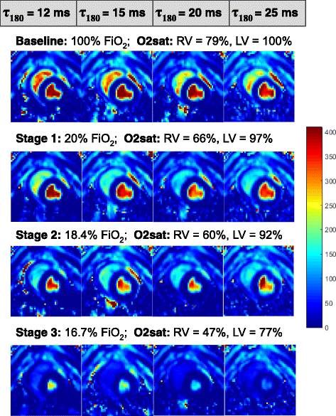
Methods: Sufficient data diversity to estimate the various unknown parameters of the L-M model, including O2 saturation, was achieved by acquiring four T2 maps, each at a different τ 180 (12, 15, 20, and 25 ms). Venous and arterial blood T2 values from these maps, together with hematocrit and arterial O2 saturation, were jointly processed to derive estimates for venous O2 saturation and other nuisance parameters in the L-M model. The technique was validated by a progressive graded hypoxemia experiment in seven pigs. CMR estimates of O2 saturation in the right ventricle were compared against a reference O2 saturation obtained by invasive catheterization from the right atrium in each pig, at each hypoxemia stage. O2 saturation derived from the proposed technique was also compared against the previously described method of applying a global calibration factor (K) to the simplified L-M model.
Results: Venous O2 saturation results obtained using the proposed CMR oximetry method exhibited better agreement (y = 0.84× + 12.29, R2 = 0.89) with invasive blood gas analysis when compared to O2 saturation estimated by a global calibration method (y = 0.69× + 27.52, R2 = 0.73).
Conclusions: We have demonstrated a novel, non-invasive method to estimate O2 saturation using quantitative T2 mapping. This technique may provide a valuable addition to the diagnostic utility of CMR in patients with congenital heart disease, heart failure, and pulmonary hypertension.
Keywords: Cardiovascular magnetic resonance; Oxygen saturation; T2 mapping.
Conflict of interest statement
Ethics approval
The study was approved by the Institutional Animal Care and Use Committee of The Ohio State University.
Consent for publication
Not Applicable.
Competing interests
JV, RA, LCP and OPS have applied for a patent relating to the contents of the manuscript.
Publisher’s Note
Springer Nature remains neutral with regard to jurisdictional claims in published maps and institutional affiliations.
Figures


Similar articles
-
Patient-Adaptive Magnetic Resonance Oximetry: Comparison With Invasive Catheter Measurement of Blood Oxygen Saturation in Patients With Cardiovascular Disease.J Magn Reson Imaging. 2020 Nov;52(5):1449-1459. doi: 10.1002/jmri.27179. Epub 2020 May 1. J Magn Reson Imaging. 2020. PMID: 32356905 Free PMC article.
-
In vivo validation of T2- and susceptibility-based Sv O2 measurements with jugular vein catheterization under hypoxia and hypercapnia.Magn Reson Med. 2019 Dec;82(6):2188-2198. doi: 10.1002/mrm.27871. Epub 2019 Jun 27. Magn Reson Med. 2019. PMID: 31250481 Free PMC article.
-
In vivo MRI measurement of blood oxygen saturation in children with congenital heart disease.Pediatr Radiol. 2005 Feb;35(2):179-85. doi: 10.1007/s00247-004-1305-6. Epub 2004 Oct 14. Pediatr Radiol. 2005. PMID: 15490150
-
Retinal oximetry and systemic arterial oxygen levels.Acta Ophthalmol. 2018 Nov;96 Suppl A113:1-44. doi: 10.1111/aos.13932. Acta Ophthalmol. 2018. PMID: 30460761 Review.
-
Estimating Coronary Sinus Oxygen Saturation from Pulmonary Artery Oxygen Saturation.Medicina (Kaunas). 2024 Nov 16;60(11):1882. doi: 10.3390/medicina60111882. Medicina (Kaunas). 2024. PMID: 39597067 Free PMC article.
Cited by
-
Value of Right and Left Ventricular T1 and T2 Blood Pool Mapping in Patients with Chronic Thromboembolic Hypertension before and after Balloon Pulmonary Angioplasty.J Clin Med. 2023 Mar 7;12(6):2092. doi: 10.3390/jcm12062092. J Clin Med. 2023. PMID: 36983095 Free PMC article.
-
2019 American Thoracic Society BEAR Cage Winning Proposal: Lung Imaging Using High-Performance Low-Field Magnetic Resonance Imaging.Am J Respir Crit Care Med. 2020 Jun 1;201(11):1333-1336. doi: 10.1164/rccm.201912-2505ED. Am J Respir Crit Care Med. 2020. PMID: 32298594 Free PMC article. No abstract available.
-
Right and left ventricular blood pool T2 ratio on cardiac magnetic resonance imaging correlates with hemodynamics in patients with pulmonary hypertension.Insights Imaging. 2023 Apr 15;14(1):66. doi: 10.1186/s13244-023-01406-9. Insights Imaging. 2023. PMID: 37060418 Free PMC article.
-
QSM Throughout the Body.J Magn Reson Imaging. 2023 Jun;57(6):1621-1640. doi: 10.1002/jmri.28624. Epub 2023 Feb 7. J Magn Reson Imaging. 2023. PMID: 36748806 Free PMC article. Review.
-
Patient-Adaptive Magnetic Resonance Oximetry: Comparison With Invasive Catheter Measurement of Blood Oxygen Saturation in Patients With Cardiovascular Disease.J Magn Reson Imaging. 2020 Nov;52(5):1449-1459. doi: 10.1002/jmri.27179. Epub 2020 May 1. J Magn Reson Imaging. 2020. PMID: 32356905 Free PMC article.
References
-
- Patel MR, Bailey SR, Bonow RO, Chambers CE, Chan PS, Dehmer GJ, Kirtane AJ, Wann LS, Ward RP. ACCF/SCAI/AATS/AHA/ASE/ASNC/HFSA/HRS/SCCM/SCCT/SCMR/STS 2012 appropriate use criteria for diagnostic catheterization: a report of the American College of Cardiology Foundation appropriate use criteria task force, Society for Cardiovascular Angiography and Interventions, American Association for Thoracic Surgery, American Heart Association, American Society of Echocardiography, American Society of Nuclear Cardiology, Heart Failure Society of America, Heart Rhythm Society, Society of Critical Care Medicine, Society of Cardiovascular Computed Tomography, Society for Cardiovascular Magnetic Resonance, and Society of Thoracic Surgeons. J Am Coll Cardiol. 2012;59(22):1995–2027. doi: 10.1016/j.jacc.2012.03.003. - DOI - PubMed
-
- Connors AF, Jr, Speroff T, Dawson NV, Thomas C, Harrell FE, Jr, Wagner D, Desbiens N, Goldman L, AW W, Califf RM, et al. The effectiveness of right heart catheterization in the initial care of critically ill patients. SUPPORT investigators. JAMA. 1996;276(11):889–897. doi: 10.1001/jama.1996.03540110043030. - DOI - PubMed
Publication types
MeSH terms
Substances
Grants and funding
- NHLBI 5T32HL098039-07/National Heart, Lung, and Blood Institute
- 15PRE24900000/American Heart Association and The Children's Heart Foundation (US)
- NIBIB R21EB021638/National Institute of Biomedical Imaging and Bioengineering
- T32 HL098039/HL/NHLBI NIH HHS/United States
- R21 EB021638/EB/NIBIB NIH HHS/United States
LinkOut - more resources
Full Text Sources
Other Literature Sources
Medical

