Intrahepatic biliary cystadenoma mimicking hydatid cyst of liver: a clinicopathologic study of six cases
- PMID: 29121977
- PMCID: PMC5680786
- DOI: 10.1186/s13256-017-1481-2
Intrahepatic biliary cystadenoma mimicking hydatid cyst of liver: a clinicopathologic study of six cases
Abstract
Background: Intrahepatic biliary cystadenomas are rare hepatic neoplasms, which are usually cystic. These tumors are often misdiagnosed as simple liver cysts and hydatid cysts clinically and radiologically owing to nonspecific clinical and radiologic features. These tumors require complete resection, as recurrence and malignant transformation can occur following incomplete excision. It is essential that these tumors be diagnosed accurately so that they can be adequately excised.
Methods: Clinical and radiological features of six cases of biliary cystadenoma are described.
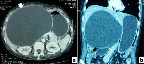
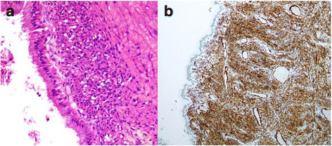
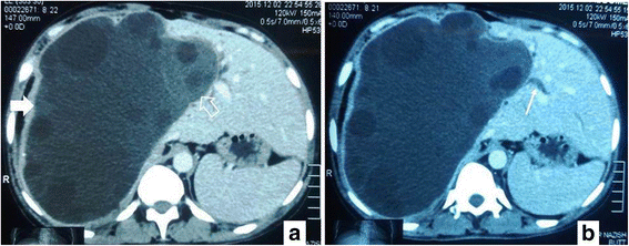
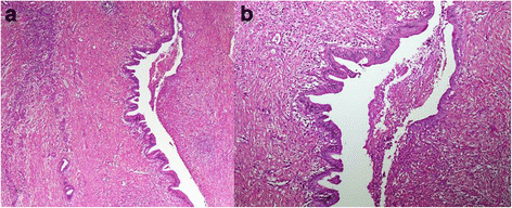
Results: All of these cases were resected with the clinical and/or radiological impression of simple liver cysts and/or hydatid cysts. Out of the six patients, five were female and one was male. Ages of the patients ranged from 28 to 60 years (mean 45 years). The patients presented with nonspecific symptoms. Internal septations were seen on preoperative imaging (when available). On gross examination, all tumors were cystic; their sizes varied from 5.5 to 14 cm, mean size was 9.0 cm. On histopathologic examination, cystic spaces were lined by cuboidal to columnar mucin-secreting epithelium with underlying ovarian-type stroma. In one case, ovarian-type stroma was not seen. Recurrence was seen in three cases at 1 to 5 years of follow up.
Conclusions: Owing to their malignant potential and high recurrence rate following incomplete resection, an aggressive surgical approach is recommended. Prognosis is excellent after complete resection.
Keywords: Biliary cystadenoma; Cyst; Hydatid cyst; Internal septations; Liver.
Conflict of interest statement
Ethics approval and consent to participate
Since this was a retrospective observational study and did not involve actual patients, patients’ images, or videos, it was granted an exemption from requiring ethics approval from the Medical Ethics Committee of Aga Khan University Hospital (4606-Pat-ERC-17).
Consent for publication
Written informed consent was obtained from close relatives of the patients (legal guardian or next of kin) for publication of the report and any accompanying images. A copy of the written consent is available for review upon request by the Editor-in-Chief of this journal.
Competing interests
The authors declare that they have no competing interests.
Publisher’s Note
Springer Nature remains neutral with regard to jurisdictional claims in published maps and institutional affiliations.
Figures


References
-
- Tsui WMS, Adsay NV, Crawford JM, et al. Mucinous cystic neoplasms of the liver. In: Bosman FT, Carneiro F, Hruban RH, Theise NE, et al., editors. WHO Classification of Tumours of the Digestive System. 4. Lyon: IARC; 2010. pp. 236–8.
-
- Weihing RR, Shintaku IP, Geller SA, Petrovic LM. Hepatobiliary and pancreatic mucinous cystadenocarcinoma with mesenchymal stroma: analysis of estrogen receptors/progesterone receptors and expression of tumor-associated antigens. Mod Pathol. 1997;10(4):372–9. - PubMed
Publication types
MeSH terms
LinkOut - more resources
Full Text Sources
Other Literature Sources
Medical

