. 2018 Jan 15:193:186-193.
doi: 10.1016/j.lfs.2017.11.002. Epub 2017 Nov 6.

CRISPR-mediated deletion of the PECAM-1 cytoplasmic domain increases receptor lateral mobility and strengthens endothelial cell junctional integrity

Affiliations

CRISPR-mediated deletion of the PECAM-1 cytoplasmic domain increases receptor lateral mobility and strengthens endothelial cell junctional integrity

Danying Liao et al. Life Sci. .

Abstract

Aims: PECAM-1 is an abundant endothelial cell surface receptor that becomes highly enriched at endothelial cell-cell junctions, where it functions to mediate leukocyte transendothelial migration, sense changes in shear and flow, and maintain the vascular permeability barrier. Homophilic interactions mediated by the PECAM-1 extracellular domain are known to be required for PECAM-1 to perform these functions; however, much less is understood about the role of its cytoplasmic domain in these processes.

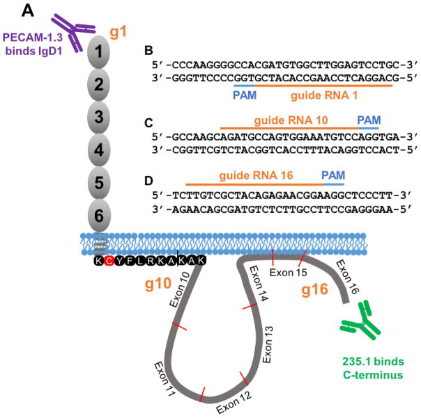
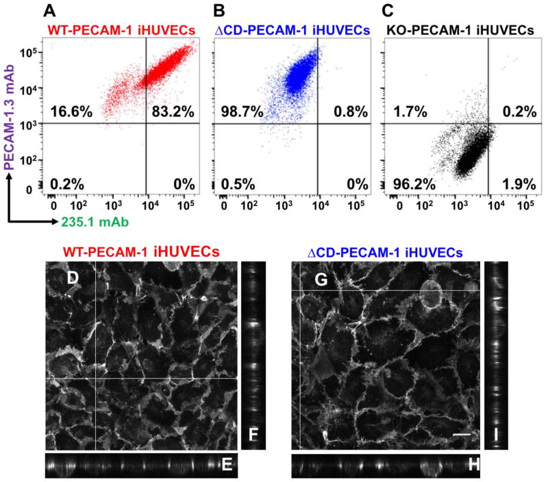
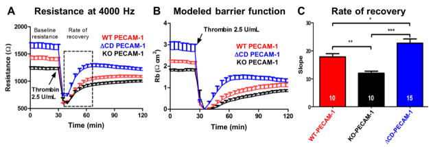
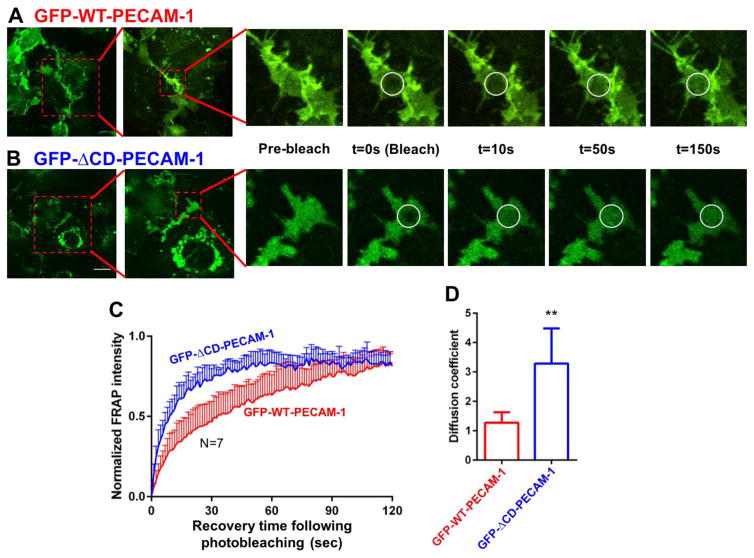
Main methods: CRISPR/Cas9 gene editing technology was employed to generate human endothelial cell lines that either lack PECAM-1 entirely, or express mutated PECAM-1 missing the majority of its cytoplasmic domain (∆CD-PECAM-1). The endothelial barrier function was evaluated by Electric Cell-substrate Impedance Sensing, and molecular mobility was assessed by fluorescence recovery after photobleaching.

Key findings: We found that ∆CD-PECAM-1 concentrates normally at endothelial cell junctions, but has the unexpected property of conferring increased baseline barrier resistance, as well as a more rapid rate of recovery of vascular integrity following thrombin-induced disruption of the endothelial barrier. Fluorescence recovery after photobleaching analysis revealed that ∆CD-PECAM-1 exhibits increased mobility within the plane of the plasma membrane, thus allowing it to redistribute more rapidly back to endothelial cell-cell borders to reform the vascular permeability barrier.

Significance: The PECAM-1 cytoplasmic domain plays a novel role in regulating the rate and extent of vascular permeability following thrombotic or inflammatory challenge.

Keywords: Adhesion; Endothelial cell; Glycosylation; PECAM-1; Permeability; Sialic acid; Vascular biology.


Conflict of interest statement

Competing interests

The authors declare no competing or financial interests.

Figures

Figure 1
Figure 1. Strategy used to generate PECAM-1 knockout and cytoplasmic domain-deleted iHUVEC cell lines
(A) Schematic of PECAM-1 showing the locations of antibody binding sites for mAb PECAM-1.3, specific for PECAM-1 IgD1, and mAb 235.1, specific for the C-terminus of the PECAM-1 cytoplasmic domain. (B) Guide RNA (gRNA) sequence (orange bar) and the protospacer adjacent motif (PAM) sequences (blue) used to introduce an insertion/deletion in exon 1 of the PECAM-1 gene to generate a PECAM-1-deficient iHUVEC line (KO-PECAM-1). (C–D) Sequence of the gRNAs that frame the PECAM-1 cytoplasmic domain used to generate an iHUVEC line expressing PECAM-1 lacking its cytoplasmic domain (ΔCD-PECAM-1). The approximate location of the binding sites of the gRNA relative to their location in exons 1, 10 and 16 are shown schematically in orange in panel A.
Figure 2
Figure 2. Characterization of CRISPR-generated iHUVEC cell lines
Flow cytometric data showing the binding of mAbs PECAM-1.3 and 235.1 to wild-type iHUVECs (panel A), ΔCD-PECAM-1 iHUVECs (panel B), and knockout PECAM-1 iHUVECs (panel C). Note the comparable surface expression levels of PECAM-1 in the WT and ΔCD iHUVEC cell lines, but absence of cytoplasmic tail in the ΔCD iHUVEC line. (D–I) Confocal fluorescence microscopy showing combined projection images (Panels D and G), as well as representative cross-sectional images (denoted by white lines) of representative z-planes (Panels E, F, H, and I) in iHUVEC cells expressing either WT-PECAM-1 or ΔCD-PECAM-1. Note that absence of the PECAM-1 cytoplasmic domain does not affect its ability to concentrate at endothelial cell-cell borders. Scale bar = 20 μm.
Figure 3
Figure 3. Absence of the PECAM-1 cytoplasmic domain confers enhanced baseline barrier function and faster restoration of endothelial cell junctional integrity following thrombin challenge
(A) ECIS analysis of the endothelial cell permeability barrier under resting and stimulated conditions of iHUVECs expressing WT and ΔCD forms of PECAM-1. The lines display the mean ± s.d. of the resistance (Ω) over time. The dashed box indicates the time frame used to calculate the rate of recovery. N=10 for the WT and KO-PECAM-1 iHUVEC lines, and 15 for the ΔCD-PECAM-1 cell line. (B) Modeled barrier function (Rb) of data shown in panel A. (C) Linear regression analysis of the resistance curves showing the mean ± s.d. of the slope from the nadir immediately after thrombin challenge to a point near full recovery. Statistics were carried out using one-way ANOVA analysis. *P<0.05; **P<0.01; *** P<0.001. Note the increased baseline barrier resistance as well as the enhanced rate of barrier recovery following thrombin stimulation of iHUVECs expressing ΔCD-PECAM-1.
Figure 4
Figure 4. Generation of iHUVEC cell lines expressing full-length and ΔCD forms of human PECAM-1 fused to GFP
Lentiviral constructs encoding fusion proteins comprised of full-length wild-type PECAM-1 fused to GFP, or ΔCD-PECAM-1 fused to GFP, were transduced into CRISPR-generated PECAM-1-negative iHUVEC cells. Transduced cells expressing similar levels of PECAM-1 were selected by fluorescence-activated cell sorting, and expression additionally evaluated by confocal microscopy (insets).
Figure 5
Figure 5. Fluorescence recovery after photobleaching (FRAP) analysis of the lateral mobility of PECAM-1 within the plane of the plasma membrane
GFP-positive cells that had formed well-defined cell-cell junctions were subjected to FRAP analysis as described in Materials and Methods. Representative images of iHUVECs expressing wild-type (panel A) and ΔCD-PECAM-1 (panel B) fused to GFP are shown at the indicated time points before and after laser-induced photobleaching. Photobleached areas are marked by white circles. (C) Normalized fluorescence intensity of GFP-WT-PECAM-1 (red) and GFP-ΔCD-PECAM-1 (blue) in the photobleached areas over time following photobleaching. (D) The diffusion coefficients for wild-type and ΔCD-PECAM-1 were calculated from the FRAP images using ImageJ. Data are expressed as the mean ± the standard deviation of seven independent experiments. Significant differences are indicated as ** P<0.01. Scale bar = 20 μm.

Similar articles

Cited by

References

    1. Albelda SM, Muller WA, Buck CA, Newman PJ. Molecular and cellular properties of PECAM-1 (endoCAM/CD31): a novel vascular cell-cell adhesion molecule. J Cell Biol. 1991;114:1059–68. - PMC - PubMed
    1. Albelda SM, Oliver PD, Romer LH, Buck CA. EndoCAM: A novel endothelial cell-cell adhesion molecule. J Cell Biol. 1990;110:1227–37. - PMC - PubMed
    1. Ayalon O, Sabanai H, Lampugnani MG, Dejana E, Geiger B. Spatial and temporal relationships between cadherins and PECAM-1 in cell-cell junctions of human endothelial cells. J Cell Biol. 1994;126:247–58. - PMC - PubMed
    1. Bergom C, Goel R, Paddock C, Gao C, Newman DK, Matsuyama S, et al. The cell-adhesion and signaling molecule PECAM-1 is a molecular mediator of resistance to genotoxic chemotherapy. Cancer Biol Ther. 2006;5:1699–707. - PubMed
    1. Biswas P, Canosa S, Schoenfeld D, Schoenfeld J, Li P, Cheas LC, et al. PECAM-1 affects GSK-3beta-mediated beta-catenin phosphorylation and degradation. Am J Pathol. 2006;169:314–24. - PMC - PubMed

MeSH terms

Substances