. 2017 Dec 15;6(12):1876-1888.
doi: 10.1242/bio.025940.

Myotube migration to cover and shape the testis of Drosophila depends on Heartless, Cadherin/Catenin, and myosin II

Affiliations

Myotube migration to cover and shape the testis of Drosophila depends on Heartless, Cadherin/Catenin, and myosin II

Silke Rothenbusch-Fender et al. Biol Open. .

Abstract

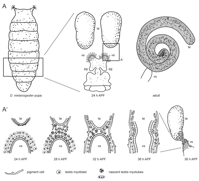
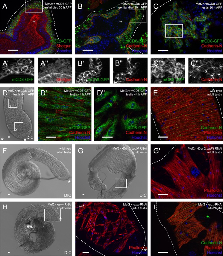
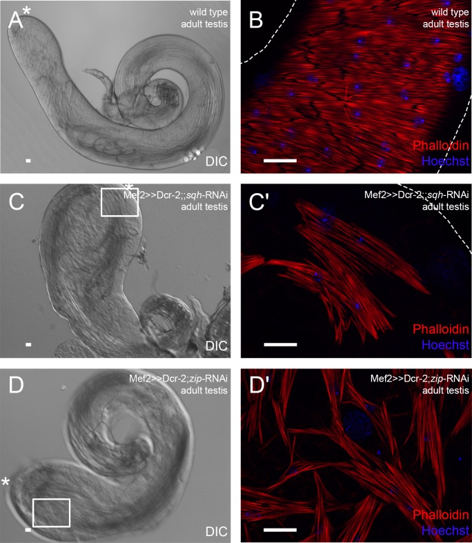
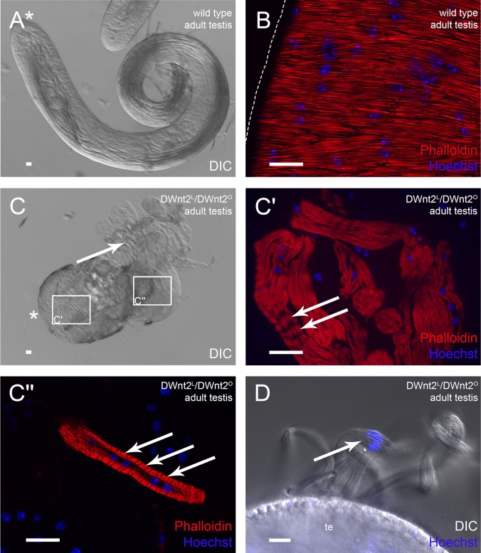
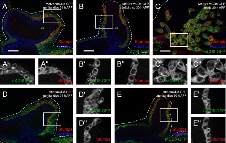
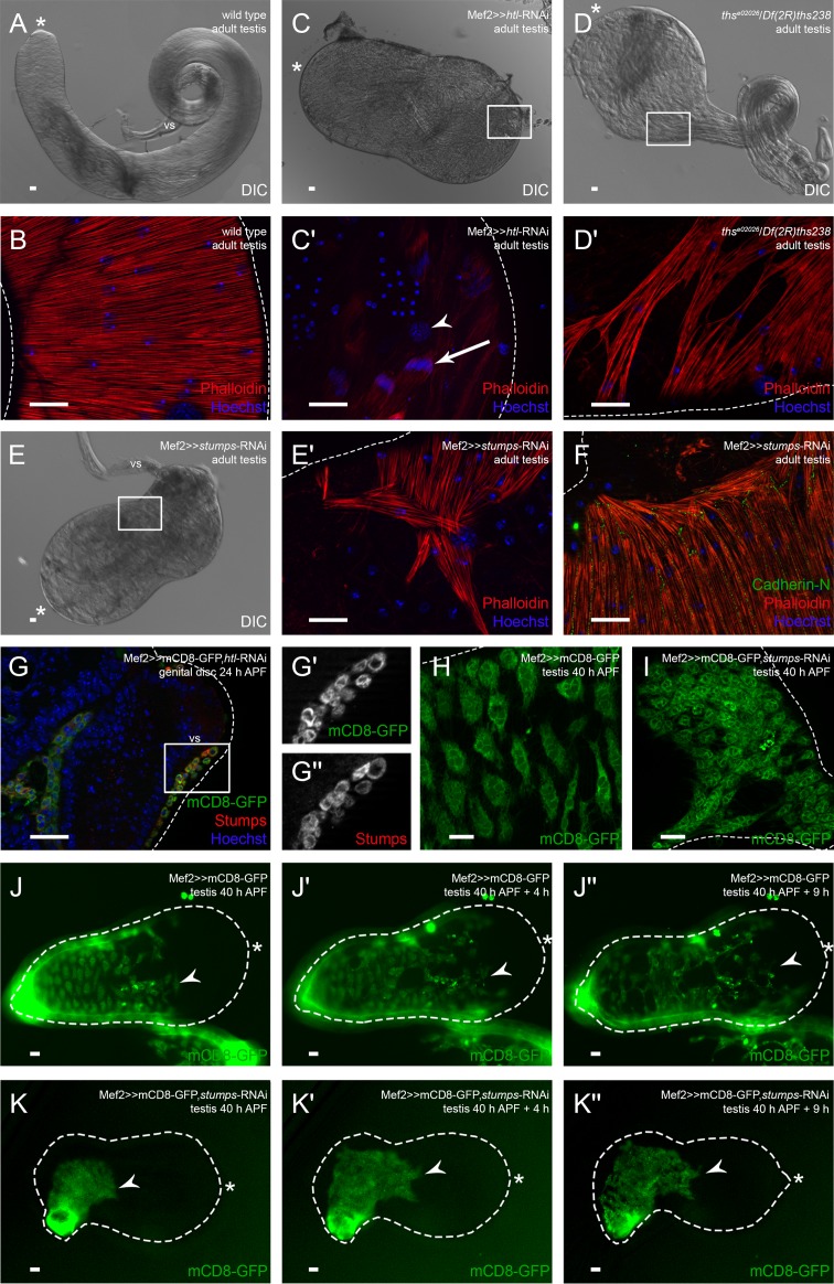
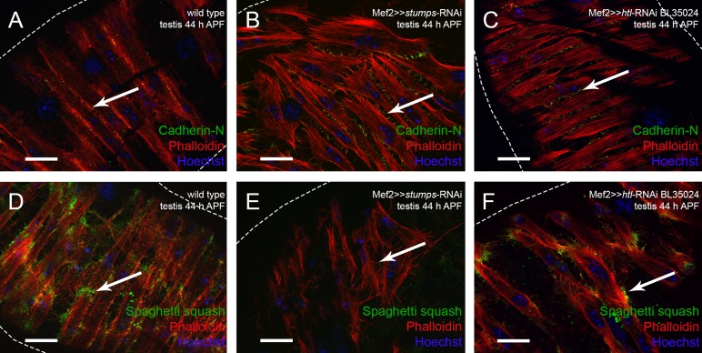
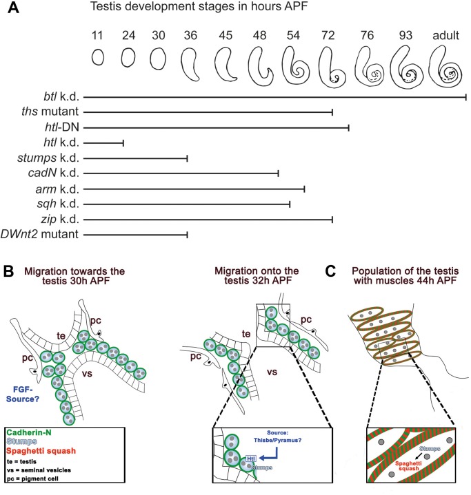
During Drosophila metamorphosis, nascent testis myotubes migrate from the prospective seminal vesicle of the genital disc onto pupal testes and then further to cover the testes with multinucleated smooth-like muscles. Here we show that DWnt2 is likely required for determination of testis-relevant myoblasts on the genital disc. Knock down of fibroblast growth factor receptor (FGFR) heartless by RNAi and a dominant-negative version revealed multiple functions of Heartless, namely regulation of the amount of myoblasts on the genital disc, connection of seminal vesicles and testes, and migration of muscles along the testes. Live imaging indicated that the downstream effector Stumps is required for migration of testis myotubes on the testis towards the apical tip. After myoblast fusion, myosin II is needed for migration of nascent testis myotubes, in which Thisbe-dependent fibroblast growth factor (FGF) signaling is activated. Cadherin-N is essential for connecting these single myofibers and for creating a firm testis muscle sheath that shapes and stabilizes the testis tubule. Based on these results, we propose a model for the migration of testis myotubes in which nascent testis myotubes migrate as a collective onto and along the testis, dependent on FGF-regulated expression of myosin II.

Keywords: DWnt2; FGF; Muscles; Stumps; Testes tubules; Thisbe.


Conflict of interest statement

Competing interestsThe authors declare no competing or financial interests.

Figures

Fig. 1.
Fig. 1.
Scheme of Drosophila testis myotube migration. (A) The male reproductive tract develops during metamorphosis. At 24 h APF, the single genital disc and paired testes (te) are separate organs. The seminal vesicles (vs) and the paragonia (pg) already start to grow. In the adult, the tubular testis is connected to the seminal vesicle. (A’) During metamorphosis, the prospective seminal vesicles and testes grow towards each other and fuse. On genital discs 24 h APF, testis-relevant myoblasts accumulate on the prospective seminal vesicle. Pigment cells cover the pupal testis. At 28 h APF, myoblasts fuse to build multinucleated testis myotubes. These nascent testis myotubes migrate beneath the pigment cells onto the pupal testis, while pigment cells migrate from the testis onto the developing seminal vesicle. By 36 h APF, the epithelia of seminal vesicles and the terminal epithelium of the testes have fused. Modified after Bodenstein (1950), Kozopas et al. (1998), Kuckwa et al. (2016).
Fig. 2.
Fig. 2.
Knock-down of Cadherin-N or Armadillo strongly reduces the adhesion between testis myotubes. Immunofluorescence analyses of genital discs and testes. (A) Seminal vesicles 30 h APF stained or marked with anti-Shotgun (red), GFP (green; myoblasts and myotubes on genital discs and pupal testes marked with Mef2≫mCD8-GFP), and Hoechst (blue; nuclei). (A′,A″) Enlargement of boxed area in A, stained or marked as indicated. (B) Genital discs 30 h APF and (C) testis 30 h APF stained or marked with anti-Cad-N (red), GFP (green), and Hoechst (blue), magnification of prospective seminal vesicle is shown. (B′,B″,C′,C″) Enlargement of boxed area in B and C stained or marked as indicated. (D–D″) Testis 44 h APF. (D) Differential interference contrast (DIC) micrograph of testis 44 h APF, (D′,D″) enlargement of boxed area in D stained or marked with anti-Cad-N (red), GFP (green), and Hoechst (blue). (E) Adult testis stained with Hoechst (blue), Phalloidin to visualize F-actin (red), and anti-Cad-N (green). (F) DIC micrograph of wild-type testis. (G) DIC micrograph of cad-N knock-down testis; (G′) enlargement of boxed area in G showing Phalloidin (red) and Hoechst (blue) staining of testis muscle sheath. (H) DIC micrograph of arm knock-down testis; (H′) enlargement of boxed area in H showing Phalloidin (red) and Hoechst (blue) staining of testis muscle sheath. (I) Adult arm knock-down testis stained with Hoechst (blue), Phalloidin to visualize F-actin (red), and anti-Cad-N (green). Dotted lines reflect approximate shape of the organ. Asterisk, hub region; vs, seminal vesicle. Scale bars: 20 µm.
Fig. 3.
Fig. 3.
Myoblast-specific down-regulation of non-muscle myosin II leads to inefficient population of the testis with muscles. Analysis of adult (A,B) wild-type testes, (C,C′) sqh knock-down testes, and (D,D′) zip knock-down testes. (A,C,D) DIC micrograph; asterisk, hub region. (B,C′,D′) Phalloidin staining to visualize F-actin (red), and Hoechst staining of nuclei (blue). C′ and D′ are enlargements of areas boxed in C and D, respectively. Dotted lines reflect approximate shape of the organ. Scale bars: 20 µm.
Fig. 4.
Fig. 4.
DWnt2 affects testis muscle determination. Analysis of adult (A,B) wild-type testes and (C-D) DWnt2L/DWnt2O testes. (A,C) DIC micrographs; arrow, leaking sperm; asterisk, hub region. (C′,C″) Enlargement of the respective boxed areas in C stained with Phalloidin to visualize F-actin (red) and Hoechst to visualize nuclei (blue). Arrows, thin actin filaments. (D) DIC micrograph and Hoechst staining (blue) of adult DWnt2L/DWnt2O testis. Arrow, nuclei of leaking spermatid bundles. Scale bars: 20 µm.
Fig. 5.
Fig. 5.
FGF signaling components are expressed during migration of testis myotubes. Immunofluorescence analysis of Stumps. Myoblasts and nascent myotubes on (A-A″) wild-type genital discs 24 h APF, (B-B″) wild-type genital discs 30 h APF (magnifications of prospective seminal vesicles are shown), and (C-C″) pupal testis 30 h APF stained or marked with anti-Stumps (red), Mef2-driven mCD8-GFP (green), and Hoechst (blue; nuclei). (A′,A″,B′,B″,C′,C″) Enlargement of boxed areas in A, B, and C, respectively, marked with GFP or stained with anti-Stumps. (D-E″) Myoblasts and myotubes on Htl-Gal4≫mCD8-GFP genital discs at (D-D″) 24 h APF (magnification of prospective seminal vesicle is shown) and (E-E″) 30 h APF, stained or marked with anti-Stumps (red), Htl-driven mCD8-GFP (green), and Hoechst (blue). (D′,D″,E′,E″) Enlargement of boxed area in D and E, respectively, marked with Htl-driven mCD8-GFP or stained with anti-Stumps. Dotted lines reflect approximate shape of the organ. vs, seminal vesicle. Scale bars: 20 µm.
Fig. 6.
Fig. 6.
Ths-activated Heartless is essential for populating the testis with myotubes. Analysis of htl and stumps knock-down and ths mutant. (A) DIC micrograph of adult wild-type testis. (B) Phalloidin staining to visualize F-actin (red), and Hoechst staining of nuclei (blue). (C) DIC micrograph of adult htl knock-down (v6692) testis. (C′) Enlargement of boxed area in C showing adult htl knock-down testis stained with Phalloidin (red; F-actin) and Hoechst (blue; nuclei); arrowhead, pigment cell nuclei; arrow, spermatids during individualization. (D) DIC micrograph of adult ths mutant testis. (D′) Enlargement of boxed area in D showing adult ths mutant testis stained with Phalloidin (red) and Hoechst (blue). (E) DIC micrograph of adult stumps knock-down testis. (E′) Enlargement of boxed area in E showing adult stumps knock-down testis stained with Phalloidin (red) and Hoechst (blue). (F) Adult stumps knock-down testis stained with anti-Cad-N (green), Phalloidin (red), and Hoechst (blue). (G-G″) htl knock-down genital disc 24 h APF stained or marked with anti-Stumps (red), GFP (green), and Hoechst (blue); magnification of prospective seminal vesicle is shown. (G′,G″) Enlargement of boxed area in G marked with GFP or stained with anti-Stumps. (H) Myoblasts on wild-type testis 40 h APF marked with Mef2-driven mCD8-GFP (green). (I) Myotubes of stumps knock-down testis 40 h APF marked with GFP (green). (J-K″) Live imaging over time of testes 40 h APF expressing Mef2-driven mCD8-GFP to reveal the migration of nascent myotubes in an ex vivo culture of (J-J″) wild-type testis and (K-K″) stumps knock-down testis. Dotted lines reflect the approximate shape of the organ. Arrowheads, the front of migrating nascent myotubes; asterisk, hub region; vs, seminal vesicle. Scale bars: 20 µm.
Fig. 7.
Fig. 7.
Expression of the myosin II subunit Spaghetti squash is not detectable after knock-down of Stumps. (A-F) Visualization of muscles on testes by Phalloidin (red). (A-C) Expression of Cad-N in (A) wild type, (B) stumps knock-down, and (C) htl knock-down. (D-F) Sqh expression in (D) wild type (Z-stacks, 27 images with 0.48 µm per layer), (E) stumps knock-down (Z-stacks, 12 images with 0.52 µm per layer), and (F) htl knock-down (Z-stacks, 9 images with 0.24 µm per layer). Arrows in A-C indicate Cad-N and in D-F indicate Sqh; dotted lines indicate border of testis.
Fig. 8.
Fig. 8.
Model of testis myotube migration. (A) Summary of shaping defects of knock-down/mutant testes. Stages of testis development in wild-type males during metamorphosis, from 11 h APF to adult. Bars indicate the stage to which to mutant testes develop [myoblast-specific knock-down (k.d.) mutants, mutant testes expressing the dominant-negative (DN) Htl protein, or a DWnt2 mutant]. According to the affected gene or genetic manipulation, testis development correlates to different stages of wild-type development. For example, down-regulation of btl does not interrupt testis development, whereas htl knock-down leads to a very early stop in development. (B,C) Two-phase model of testis myotube migration. (B) Phase one represents the Htl dependent migration of nascent myotubes onto the testis before and after fusion of the epithelium of the seminal vesicle (vs) and the terminal epithelium (te) of the testes. Pigment cells (pc) migrated towords the vs. The FGF ligand Ths and possibly Pyr are secreted by an unknown source. (C) In phase two, nascent myotubes are already on the testis and migrate further towards the tip. Cad-N mediates adhesion between adjacent myotubes. Stumps might control the migration process, likely via non-muscle myosin II. Sqh expression depends on Stumps, which suggests Sqh regulation by Htl signaling. Note that after an initial phase one, both phases run in parallel until all myotubes reach the testis.

Similar articles

Cited by

References

    1. Ahmad S. M. and Baker B. S. (2002). Sex-specific deployment of FGF signaling in Drosophila recruits mesodermal cells into the male genital imaginal disc. Cell 109, 651-661. 10.1016/S0092-8674(02)00744-4 - DOI - PubMed
    1. Aldaz S., Escudero L. M. and Freeman M. (2010). Live imaging of Drosophila imaginal disc development. Proc. Natl. Acad. Sci. USA 107, 14217-14222. 10.1073/pnas.1008623107 - DOI - PMC - PubMed
    1. Awe S. and Renkawitz-Pohl R. (2010). Histone H4 acetylation is essential to proceed from a histone- to a protamine-based chromatin structure in spermatid nuclei of Drosophila melanogaster. Syst. Biol. Reprod. Med. 56, 44-61. 10.3109/19396360903490790 - DOI - PubMed
    1. Baylies M. K., Martinez Arias A. and Bate M. (1995). wingless is required for the formation of a subset of muscle founder cells during Drosophila embryogenesis. Development 121, 3829-3837. - PubMed
    1. Bertrand V. (2016). Beta-catenin-driven binary cell fate decisions in animal development. Wiley Interdiscip. Rev. Dev. Biol. 5, 377-388. 10.1002/wdev.228 - DOI - PMC - PubMed

LinkOut - more resources