

Mechanism of polypurine tract primer generation by HIV-1 reverse transcriptase
- PMID: 29122886
- PMCID: PMC5766924
- DOI: 10.1074/jbc.M117.798256
Mechanism of polypurine tract primer generation by HIV-1 reverse transcriptase
Abstract
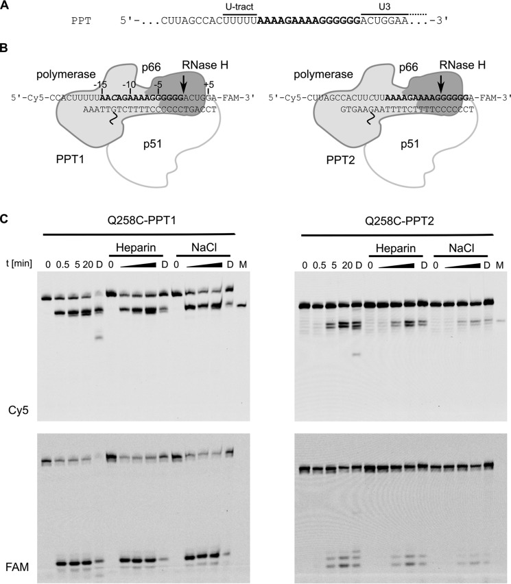
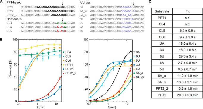
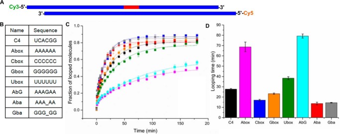
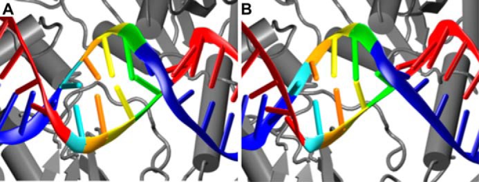
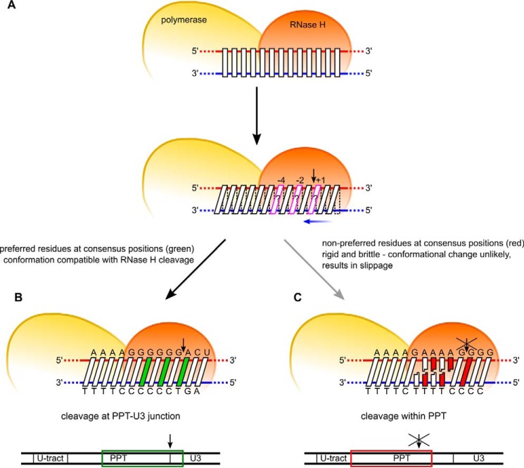
HIV-1 reverse transcriptase (RT) possesses both DNA polymerase activity and RNase H activity that act in concert to convert single-stranded RNA of the viral genome to double-stranded DNA that is then integrated into the DNA of the infected cell. Reverse transcriptase-catalyzed reverse transcription critically relies on the proper generation of a polypurine tract (PPT) primer. However, the mechanism of PPT primer generation and the features of the PPT sequence that are critical for its recognition by HIV-1 RT remain unclear. Here, we used a chemical cross-linking method together with molecular dynamics simulations and single-molecule assays to study the mechanism of PPT primer generation. We found that the PPT was specifically and properly recognized within covalently tethered HIV-1 RT-nucleic acid complexes. These findings indicated that recognition of the PPT occurs within a stable catalytic complex after its formation. We found that this unique recognition is based on two complementary elements that rely on the PPT sequence: RNase H sequence preference and incompatibility of the poly(rA/dT) tract of the PPT with the nucleic acid conformation that is required for RNase H cleavage. The latter results from rigidity of the poly(rA/dT) tract and leads to base-pair slippage of this sequence upon deformation into a catalytically relevant geometry. In summary, our results reveal an unexpected mechanism of PPT primer generation based on specific dynamic properties of the poly(rA/dT) segment and help advance our understanding of the mechanisms in viral RNA reverse transcription.
Keywords: cysteine-mediated cross-linking; human immunodeficiency virus (HIV); molecular dynamics; nucleic acid structure; protein-nucleic acid interaction; reverse transcriptase; ribonuclease H.
© 2018 by The American Society for Biochemistry and Molecular Biology, Inc.
Conflict of interest statement
The authors declare that they have no conflicts of interest with the contents of this article
Figures
Similar articles
-
Pre-existing distortions in nucleic acid structure aid polypurine tract selection by HIV-1 reverse transcriptase.J Biol Chem. 2002 May 10;277(19):16689-96. doi: 10.1074/jbc.M109914200. Epub 2002 Mar 1. J Biol Chem. 2002. PMID: 11875059
-
Two modes of HIV-1 polypurine tract cleavage are affected by introducing locked nucleic acid analogs into the (-) DNA template.J Biol Chem. 2004 Aug 27;279(35):37095-102. doi: 10.1074/jbc.M403306200. Epub 2004 Jun 25. J Biol Chem. 2004. PMID: 15220330
-
Combining mutations in HIV-1 reverse transcriptase with mutations in the HIV-1 polypurine tract affects RNase H cleavages involved in PPT utilization.Virology. 2006 May 10;348(2):378-88. doi: 10.1016/j.virol.2005.12.042. Epub 2006 Feb 10. Virology. 2006. PMID: 16473384
-
'Binding, bending and bonding': polypurine tract-primed initiation of plus-strand DNA synthesis in human immunodeficiency virus.Int J Biochem Cell Biol. 2004 Sep;36(9):1752-66. doi: 10.1016/j.biocel.2004.02.016. Int J Biochem Cell Biol. 2004. PMID: 15183342 Review.
-
Interaction of retroviral reverse transcriptase with template-primer duplexes during replication.Prog Nucleic Acid Res Mol Biol. 1998;58:339-93. doi: 10.1016/s0079-6603(08)60041-0. Prog Nucleic Acid Res Mol Biol. 1998. PMID: 9308371 Review.
Cited by
-
GC-Content Dependence of Elastic and Overstretching Properties of DNA:RNA Hybrid Duplexes.Biophys J. 2020 Aug 18;119(4):852-861. doi: 10.1016/j.bpj.2020.06.034. Epub 2020 Jul 16. Biophys J. 2020. PMID: 32738216 Free PMC article.
-
HIV-1 Reverse Transcriptase Inhibition by Major Compounds in a Kenyan Multi-Herbal Composition (CareVid™): In Vitro and In Silico Contrast.Pharmaceuticals (Basel). 2021 Sep 30;14(10):1009. doi: 10.3390/ph14101009. Pharmaceuticals (Basel). 2021. PMID: 34681233 Free PMC article.
-
Structural basis of R-loop recognition by the S9.6 monoclonal antibody.Nat Commun. 2022 Mar 28;13(1):1641. doi: 10.1038/s41467-022-29187-7. Nat Commun. 2022. PMID: 35347133 Free PMC article.
-
Role of salt-bridging interactions in recognition of viral RNA by arginine-rich peptides.Biophys J. 2021 Nov 16;120(22):5060-5073. doi: 10.1016/j.bpj.2021.10.007. Epub 2021 Oct 26. Biophys J. 2021. PMID: 34710377 Free PMC article.
-
Comprehensive Assessment of Force-Field Performance in Molecular Dynamics Simulations of DNA/RNA Hybrid Duplexes.J Chem Theory Comput. 2024 Aug 13;20(15):6917-6929. doi: 10.1021/acs.jctc.4c00601. Epub 2024 Jul 16. J Chem Theory Comput. 2024. PMID: 39012172 Free PMC article.
References
-
- Telesnitsky A., and Goff S. P. (1997) Reverse transcriptase and the generation of retroviral DNA, in Retroviruses (Coffin J. M., Hughes S. H., and Varmus H. E., eds), pp. 121–160, Cold Spring Harbor Laboratory Press, Cold Spring Harbor, NY - PubMed
-
- Le Grice S. F. J., and Nowotny M. (2014) Reverse transcriptases, in Nucleic Acid Polymerases (Murakami K., and Trakselis M. A., eds) pp. 189–214, Springer-Verlag, Berlin
Publication types
MeSH terms
Substances
Associated data
- Actions
- Actions
Grants and funding
LinkOut - more resources
Full Text Sources
Other Literature Sources
