. 2017 Dec 29;292(52):21490-21503.
doi: 10.1074/jbc.RA117.000113. Epub 2017 Nov 9.

The cell adhesion molecule BT-IgSF is essential for a functional blood-testis barrier and male fertility in mice

Affiliations

The cell adhesion molecule BT-IgSF is essential for a functional blood-testis barrier and male fertility in mice

Laura Pelz et al. J Biol Chem. .

Abstract

The Ig-like cell adhesion molecule (IgCAM) BT-IgSF (brain- and testis-specific Ig superfamily protein) plays a major role in male fertility in mice. However, the molecular mechanism by which BT-IgSF supports fertility is unclear. Here, we found that it is localized in Sertoli cells at the blood-testis barrier (BTB) and at the apical ectoplasmic specialization. The absence of BT-IgSF in Sertoli cells in both global and conditional mouse mutants (i.e. AMHCre and Rosa26CreERT2 lines) resulted in male infertility, atrophic testes with vacuolation, azoospermia, and spermatogenesis arrest. Although transcripts of junctional proteins such as connexin43, ZO-1, occludin, and claudin11 were up-regulated in the absence of BT-IgSF, the functional integrity of the BTB was impaired, as revealed by injection of a BTB-impermeable component into the testes under in vivo conditions. Disruption of the BTB coincided with mislocalization of connexin43, which was present throughout the seminiferous epithelium and not restricted to the BTB as in wild-type tissues, suggesting impaired cell-cell communication in the BT-IgSF-KO mice. Because EM images revealed a normal BTB structure between Sertoli cells in the BT-IgSF-KO mice, we conclude that infertility in these mice is most likely caused by a functionally impaired BTB. In summary, our results indicate that BT-IgSF is expressed at the BTB and is required for male fertility by supporting the functional integrity of the BTB.

Keywords: BT-IgSF (IgSF11); IgCAM; IgCAM BT-IgSF (IgSF11); blood–testis barrier; cell adhesion; connexin43; male fertility; spermatogenesis; testis.


Conflict of interest statement

The authors declare that they have no conflicts of interest with the contents of this article

Figures

Figure 1.
Figure 1.
BT-IgSF knock-out males reveal a disturbed testis morphology and are infertile. A, immunofluorescence staining of wild-type and BT-IgSF knock-out testis sections with a BT-IgSF antibody. Arrow, expression at the adluminal side, in elongated spermatids; arrowheads, expression at the basal side, in Sertoli cells; scale bar, 50 μm. B, immunofluorescence staining of wild-type testis sections against BT-IgSF. Arrows, expression at elongated spermatids at the apical ES; arrowhead, expression at the basal side (marked by white dashed line), in Sertoli cells (indicated by yellow dashed line); scale bar, 5 μm. C, immunofluorescence staining of epididymis sections show labeling of mature sperm. Scale bar, 50 μm. D, immunofluorescence staining of testis sections show BT-IgSF co-localized with Espin at the apical ES. Scale bar, 10 μm. E, immunofluorescence staining of wild-type testis sections against BT-IgSF and mouse anti-Cx43; Cx43 co-localizes partly with BT-IgSF. Arrow, specific expression at the basal side (please compare with staining of rabbit anti-Cx43 Fig. 4F); arrowheads, unspecific staining of a band around the tubuli by the secondary goat anti-mouse A488 antibody (see also S2F); scale bar, 25 μm. F, immunofluorescence staining of wild-type testis sections against BT-IgSF and mouse anti-ZO-1. Note the strong co-localization of BT-IgSF and ZO-1. Arrows, specific expression at the basal side (please compare with staining of rabbit anti-ZO-1 Fig. 4E); arrowheads, unspecific staining of a band around the tubuli by the secondary goat anti-mouse A488 antibody (see also S2F); scale bar, 20 μm. G, BT-IgSF knock-out animals exhibit smaller testes. bl, bladder; e, epididymis; f, fat; p, penis; pg, preputial gland; sv, seminal vesicle; t, testis; scale bar, 5 mm. H, knock-out testes showed a significantly decreased wet weight compared with wild-type and heterozygous testes; p value < 0.001 (one-way analysis of variance with Bonferroni correction; WT animals, n = 14; heterozygous animals, n = 22; KO animals, n = 30). The data are shown as means ± S.D. I, H&E staining of methacrylate-sections show atrophic testes of BT-IgSF knock-out animals. Black arrow, multinucleated cells; arrowheads, Sertoli cell cytoplasmic extensions; asterisk, vacuolar degeneration; scale bar, 100 μm. Insets shows the lack of sperms in KO animals at higher magnification (blue arrow). J, flow cytometric analysis of testis cells, stained with DAPI, shows the lack of haploid chromatin containing cells in BT-IgSF knockouts (red). 1c, haploid, 23 chromatids; 2c, haploid, 23 chromosomes; S, S phase; 4c, diploid, 46 chromosomes.
Figure 2.
Figure 2.
The lack of BT-IgSF leads to spermatogenic arrest. A, qRT-PCR analysis of meiotic genes from testis lysates of wild-type and BT-IgSF knock-out mice. Gene expression was normalized to Rplp0 and indicated as fold change to WT. Piwil2 was used as a marker for stem cells, HoxA4 and Cdc25c were used as markers for mid-pachytene stage and late pachytene stage, for overall pachytene stage SycP3 was used, for meiotic germ cells Dazl was used, and for the postmeiotic haploid stages Prm1 and Akap4 were used (n = 4 per genotype). The data are shown as means ± S.D. n.s., not significant; **, p < 0.01; ***, p < 0.001 (t test). The scheme illustrates the pattern of expression of genes investigated during germ maturation. B, immunofluorescence staining of cryosections against γH2AX and SYCP3. Arrows, leptotene stage; arrowheads, diplotene stage; scale bar, 20 μm.
Figure 3.
Figure 3.
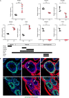
Loss of BT-IgSF influences the overall morphology of Sertoli cells but not the hormonal balance. A, immunofluorescence staining against vimentin, a cytoplasmic Sertoli cell marker. Note the irregular and disturbed cytoplasmic arms of Sertoli cells in BT-IgSF knock-out mice. Scale bar, 50 μm. B, immunofluorescence staining against WT1, a Sertoli cell marker located in the nucleus, shows normal Sertoli cell localization in tubuli and number in knock-out mice. Scale bar, 50 μm. C, immunofluorescence staining against active caspase-3, a marker for apoptosis, shows slightly more apoptotic cells in knock-out testes, especially in Sertoli cells (inset). Scale bar, 50 μm. D, quantitation of apoptotic cells per tubuli. No significant difference between knock-out and wild-type testes was observed (n = 5 per group). The data are shown as means ± S.D. (p = 0.08; t test). E, no hormonal changes in LH (p = 0.78), FSH (p = 0.69), and testosterone (p = 0.79) levels were detected in BT-IgSF knock-out males, compared with wild-type (WT, n = 5; KO, n = 6). The data are shown as means ± S.D. (differences were not significant; Mann–Whitney test).
Figure 4.
Figure 4.
BT-IgSF loss leads to an impaired BTB and up-regulation of transcripts encoding BTB proteins. A, in vivo biotin assay to test the functionality of the BTB. Biotin distribution is visualized by streptavidin-Cy5 staining; knock-out testis show distribution through all layers of the seminiferous epithelium, indicating an impaired BTB (n = 3 per genotype). Scale bar, 50 μm. B, analysis of BTB gene expression by qRT-PCR; Gja1, Tjp1, Occludin, and Cldn11 are up-regulated in knock-out testes (n = 4 per group). The data are shown as means ± S.D. *, p < 0.05 (t test). C and D, protein expression of Cx43. P0, unphosphorylated; P1 and P2, phosphorylated forms. Anti-pan-cadherin was used as a loading control. Three independent blots were analyzed that showed no significant differences between WT and KO (t test). The data are shown as means ± S.D. E, immunofluorescence staining against ZO-1, a tight junction protein of the BTB; note the slightly less organized localization of ZO-1 in knock-out testis. Scale bar, 50 μm. F, immunofluorescence staining against Cx43, a gap junction protein of the BTB; note the diffuse localization of Cx43 in knock-out testis. Scale bar, 20 μm. G, electron microscopy of adult testis. At higher magnifications, no differences between WT and KO were observed in the basal ES, typified by the actin filament bundles (blue arrowheads) sandwiched between cisternae of the endoplasmic reticulum (green arrowheads) and the plasma membranes of two Sertoli cells, tight junction (white arrowheads) co-existing with the basal ES. Red outlines, vacuoles; S, Sertoli cells; scale bars, 10 μm for left panel (WT and KO) and 500 nm for right panel (WT and KO).
Figure 5.
Figure 5.
Infertility is due to the absence of BT-IgSF in Sertoli cells. A, AMH-cKO testes show a significantly decreased wet weight compared with control animals (n = 3 for control and n = 4 for AMH-cKO). The data are shown as means ± S. D. ***, p < 0.001 (t test). B, H&E stainings of methacrylate-sections show atrophic testes of AMH-cKO mice compared with wild type. Scale bar, 50 μm. C, immunofluorescence staining against BT-IgSF in AMH-cKO showing the absence of BT-IgSF signal in Sertoli cells (arrowhead). Scale bar, 50 μm. D, flow cytometric analysis of testis cells, stained with DAPI, shows the lack of haploid chromatin containing cells in AMH-conditional knockouts (red) compared with BT-IgSFflx/flx control (black). 1c, haploid, 23 chromatids; 2c, haploid, 23 chromosomes; S, S phase; 4c, diploid, 46 chromosomes.
Figure 6.
Figure 6.
Loss of BT-IgSF in adulthood also affects spermatogenesis. A, H&E stainings of methacrylate-sections show atrophic testes of BT-IgSFflx/flx;Rosa26CreERT2 mice treated with tamoxifen compared with vehicle control. B, BT-IgSFflx/flx;Rosa26CreERT2 testis treated with tamoxifen show a significantly decreased wet weight compared with vehicle treated animals (n = 4 for vehicle and n = 4 for tamoxifen). The data are shown as means ± S.D. ***, p < 0.001 (t test). C and D, flow cytometric analysis of testis cells, stained with DAPI, shows the reduction of haploid chromatin containing cells by tamoxifen treatment of BT-IgSFflx/flx;Rosa26CreERT2 (red) compared with vehicle control (black) and the increase in 2C and 4C cells. 1c, haploid, 23 chromatids; 2c, haploid, 23 chromosomes; S, S phase; 4c, diploid, 46 chromosomes. E, analysis of expression of Cx43 and Prm1 transcripts by qRT-PCR. BT-IgSF expression is reduced by ∼50% in tamoxifen-treated testes. Gja1 (Cx43) is up-regulated in tamoxifen treated testes; Prm1, a marker for spermatids and sperm, is significantly reduced (n = 4 per group). The data are shown as means ± S.D. *, p < 0.05; **, p < 0.01; ***, p < 0.001 (t test).

Similar articles

Cited by

References

    1. Suzu S., Hayashi Y., Harumi T., Nomaguchi K., Yamada M., Hayasawa H., and Motoyoshi K. (2002) Molecular cloning of a novel immunoglobulin superfamily gene preferentially expressed by brain and testis. Biochem. Biophys. Res. Commun. 296, 1215–1221 - PubMed
    1. Katoh M., and Katoh M. (2003) IGSF11 gene, frequently up-regulated in intestinal-type gastric cancer, encodes adhesion molecule homologous to CXADR, FLJ22415 and ESAM. Int. J. Oncol. 23, 525–531 - PubMed
    1. Matthäus C., Langhorst H., Schütz L., Jüttner R., and Rathjen F. G. (2017) Cell–cell communication mediated by the CAR subgroup of immunoglobulin cell adhesion molecules in health and disease. Mol. Cell. Neurosci. 81, 32–40 - PubMed
    1. Eom D. S., Inoue S., Patterson L. B., Gordon T. N., Slingwine R., Kondo S., Watanabe M., and Parichy D. M. (2012) Melanophore migration and survival during zebrafish adult pigment stripe development require the immunoglobulin superfamily adhesion molecule Igsf11. PLoS Genet. 8, e1002899. - PMC - PubMed
    1. Harada H., Suzu S., Hayashi Y., and Okada S. (2005) BT-IgSF, a novel immunoglobulin superfamily protein, functions as a cell adhesion molecule. J. Cell. Physiol. 204, 919–926 - PubMed

Publication types

MeSH terms