. 2018 Jan 26;293(4):1151-1162.
doi: 10.1074/jbc.RA117.000160. Epub 2017 Nov 9.

Phenotypic dissection of the mouse Ren1d knockout by complementation with human renin

Affiliations

Phenotypic dissection of the mouse Ren1d knockout by complementation with human renin

Charlotte Buckley et al. J Biol Chem. .

Abstract

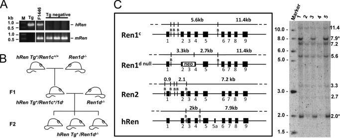
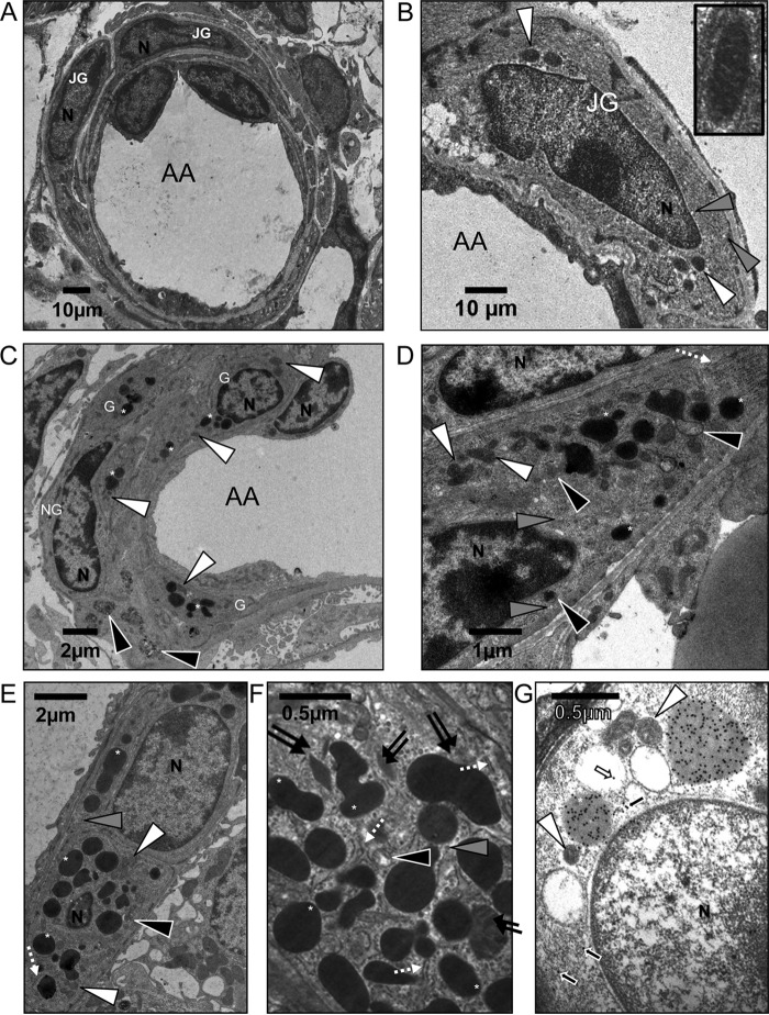
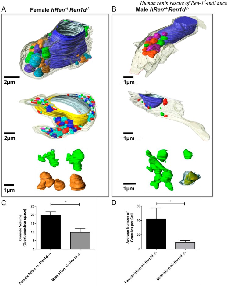
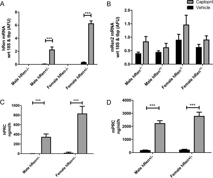
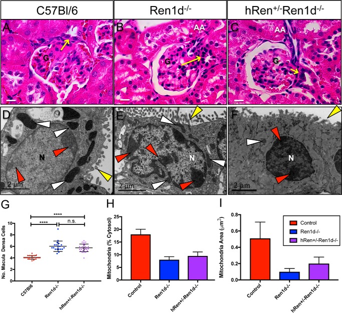
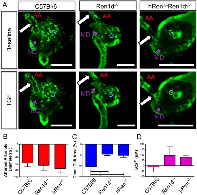
Normal renin synthesis and secretion is important for the maintenance of juxtaglomerular apparatus architecture. Mice lacking a functional Ren1d gene are devoid of renal juxtaglomerular cell granules and exhibit an altered macula densa morphology. Due to the species-specificity of renin activity, transgenic mice are ideal models for experimentally investigating and manipulating expression patterns of the human renin gene in a native cellular environment without confounding renin-angiotensin system interactions. A 55-kb transgene encompassing the human renin locus was crossed onto the mouse Ren1d-null background, restoring granulation in juxtaglomerular cells. Correct processing of human renin in dense core granules was confirmed by immunogold labeling. After stimulation of the renin-angiotensin system, juxtaglomerular cells contained rhomboid protogranules with paracrystalline contents, dilated rough endoplasmic reticulum, and electron-lucent granular structures. However, complementation of Ren1d-/- mice with human renin was unable to rescue the abnormality seen in macula densa structure. The juxtaglomerular apparatus was still able to respond to tubuloglomerular feedback in isolated perfused juxtaglomerular apparatus preparations, although minor differences in glomerular tuft contractility and macula densa cell calcium handling were observed. This study reveals that the human renin protein is able to complement the mouse Ren1d-/- non-granulated defect and suggests that granulopoiesis requires a structural motif that is conserved between the mouse Ren1d and human renin proteins. It also suggests that the altered macula densa phenotype is related to the activity of the renin-1d enzyme in a local juxtaglomerular renin-angiotensin system.

Keywords: animal model; confocal microscopy; electron microscopy (EM); granulation; human renin; immunochemistry; juxtaglomerular; macula densa; mouse; renin; renin angiotensin system; secretion; transgenic mice.


Conflict of interest statement

The authors declare that they have no conflicts of interest with the contents of this article

Figures

Figure 1.
Figure 1.
A, transgene positive animals were identified by PCR. Marker (M), New England Biolabs 1-kb DNA Ladder; Tg, PAC 111L11; F1446, hRen+; Tg negative: transgenic negative animals. B, breeding strategy to move the hRen transgene from a Ren1c background to the Ren1d −/− background. C, the structure of the mouse and human renin genes and the predicted sizes of BamHI fragments that are detected by a mouse Ren2 cDNA hybridization probe are shown in the left panel. The autoradiograph (right) is an exemplary Southern blot showing the genotyping of F1 backcross progeny. Inheritance of the human renin transgene is indicated by the presence of diagnostic human-specific fragments of 2.0 and 7.9 kb (asterisks).
Figure 2.
Figure 2.
Ultrastructure of kidney juxtaglomerular cells from hRen+/−Ren1d−/− and Ren1d−/− mice. For all images: G, granulated JG cell; NG, non-granulated JG cell; N, nucleus; white arrowhead, mitochondria; gray arrowhead, electron-lucent vesicle; black arrowhead, immature renin-containing granules; dashed white arrow, rough endoplasmic reticulum; double black arrow, paracrystalline protogranule; white asterisk, dense core renin-containing granule. A, low magnification image of afferent arteriole surrounded by juxtaglomerular cells in a Ren1d−/− mouse, showing no electron dense granules. B, high magnification juxtaglomerular cells from Ren1d−/− mice, containing no granules. Inset shows high magnification image of mitochondria. C, low magnification, sparse granulation of male hRen+/−Ren1d−/− juxtaglomerular cells surrounding the afferent arteriole. G, granulated JG cells; NG, non-granulated JG cell. D, high magnification ultrastructure of male hRen+/−Ren1d−/− juxtaglomerular cells showing sparse granulation. E and F, highly granulated female hRen+/−Ren1d−/− juxtaglomerular cell. G, anti-hRen immunogold-labeled sections from male hRen+/−Ren1d−/− juxtaglomerular cells. White arrows, immunogold labeling within electron-lucent granules; black arrows, immunogold labeling within cytoplasm. Scale bars are represented individually.
Figure 3.
Figure 3.
3D reconstruction of juxtaglomerular cells from hRen-complemented Ren1d−/− mice. Reconstructions of serial electron micrographs were rendered using Amira (FEI) software for female (A) and male (B) hRen+/−Ren1d−/− juxtaglomerular cells. The nucleus is represented in blue or yellow. Granules are colored to allow ease of differentiation between solitary or networked granules. C, the volume of the rendered granules was assessed with respect to the percentage of extranuclear space and compared between female and male hRen+/−Ren1d−/− mice. D, the average number of granules was also assessed and compared between male and female hRen+/−Ren1d−/− animals. Error bars are mean ± S.E. from cells with n > 3 in each group. *, p < 0.05; **, p < 0.01 by Student's t test. Scale bars are represented individually.
Figure 4.
Figure 4.
A and B, whole kidney assessment of human renin (A) and mouse renin (B) transcript levels in male and female hRen+/−Ren1d−/− and hRen−/− Ren1d−/− untreated or captopril-treated (1 mg/ml in the drinking water for 10 days, n = 6) mice. C, human plasma renin concentration (hPRC), and D, mouse plasma renin concentration (mPRC) in response to ACE inhibition with captopril or treatment with vehicle in male and female hRen+/−Ren1d−/− mice (n = 3–7). Error bars represent S.E. with *, p < 0.05; **, p < 0.01; ***, p < 0.001; ****, p < 0.0001 by two-way analysis of variance in conjunction with Bonferroni post hoc analysis.
Figure 5.
Figure 5.
Ultrastructure of juxtaglomerular cells in Ren1d−/− and hRen+/−Ren1d−/− mice after ACE inhibition. Animals were administered captopril (1 mg/ml in drinking water for 10 days). For all images, N, nucleus; white arrowheads, mitochondria; gray arrowheads, electron-lucent vesicles; black arrowheads, electron-lucent granules; single black arrow, electron-lucent lysosomes; white arrows, granule merging; dashed white arrow, rough endoplasmic reticulum; double black arrow, protogranules containing paracrystalline material; EC, endothelial cells; black star, gap junction; white asterisk, dense core renin-containing granule. Scale bars represent 500 nm. A–C, Ren1d−/− mice. D–F, hRen+/−Ren1d−/− male mice. G, granule lucency was measured in 8 cells from 8 different mice per group, where the mean pixel intensity was analyzed with respect to the electron density of the cytosol. ****, p < 0.0001 by Student's t test. Error bars represent S.E.
Figure 6.
Figure 6.
Macula densa morphology remains altered in hRen rescue mice on a Ren1d−/− background. A–C, H&E-stained kidneys sections showing the macula densa at its JGA position from male C57Bl/6, Ren1d−/−, and hRen+/−Ren1d−/− mice. Yellow arrow, macula densa plaque; N, nucleus; G, glomerular tuft. Scale bars represent 20 μm. D–F, electron micrographs of macula densa cells in C57Bl/6, Ren1d−/−, and hRen+/−Ren1d−/− mice. N, nucleus; white arrowheads, mitochondria; red arrowheads, dense packed chromatin; yellow arrowheads, microvilli. Scale bars represent 2 μm. G, quantification of macula densa cell number in a plaque (n = 4 Ren1d−/−; n = 5 C57Bl/6, hRen+/−Ren1d−/−). ****, = p < 0.0001. H and I, quantification of a cytoplasmic area occupied by mitochondria, and mitochondrial area in C57Bl/6, Ren1d−/−, and hRen+/−Ren1d−/− mice.
Figure 7.
Figure 7.
Tubuloglomerular feedback functions appropriately in isolated perfused juxtaglomerular apparatus from C57Bl/6, Ren1d−/−, and hRen+/−Ren1d−/− mice. Flow-induced TGF was triggered by increasing the rate of constant 10 mm NaCl-containing tubular perfusion from 2 to 20 nl/min. A, TGF is manifested by constriction of the glomerular tuft area and afferent arteriole diameter in C57Bl/6, Ren1d−/−, and hRen+/−Ren1d−/− mice. G, glomerulus. Glomerular architecture was visualized and assessed using ratiometric calcium indicator dyes Fluo-4 (green) and Fura Red (red, not shown). Scale bars represent 100 μm. White arrows indicate the location of AA constriction at baseline and after triggering TGF. TGF response was quantified by measuring the change in (B) AA diameter, (C) glomerular tuft area, and (D) calcium concentration at the macula densa plaque. Error bars = S.E., *, p < 0.05; **, p < 0.01 using Student's t test. n > 3 in each group.

Similar articles

Cited by

References

    1. Hackenthal E., Paul M., Ganten D., and Taugner R. (1990) Morphology, physiology, and molecular biology of renin secretion. Physiol. Rev. 70, 1067–1116 - PubMed
    1. Steppan D., Zügner A., Rachel R., and Kurtz A. (2013) Structural analysis suggests that renin is released by compound exocytosis. Kidney Int. 83, 233–241 - PubMed
    1. Ogawa K., Yamasato M., and Taniguchi K. (1995) Exocytosis of secretory granules in the juxtaglomerular granular cells of kidneys. Anat. Rec. 243, 336–346 10.1002/ar.1092430308 - DOI - PubMed
    1. Panthier J. J., and Rougeon F. (1983) Kidney and submaxillary gland renins are encoded by two non-allelic genes in swiss mice. EMBO J. 2, 675–678 - PMC - PubMed
    1. Mullins J. J., Burt D. W., Windass J. D., McTurk P., George H., and Brammar W. J. (1982) Molecular cloning of two distinct renin genes from the dba/2 mouse. EMBO J. 1, 1461–1466 - PMC - PubMed

Publication types

LinkOut - more resources