. 2017 Nov 10;358(6364):813-818.
doi: 10.1126/science.aao3265.

SAMTOR is an S-adenosylmethionine sensor for the mTORC1 pathway

Affiliations

SAMTOR is an S-adenosylmethionine sensor for the mTORC1 pathway

Xin Gu et al. Science. .

Abstract

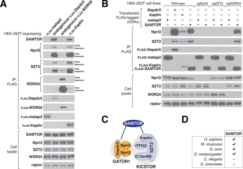
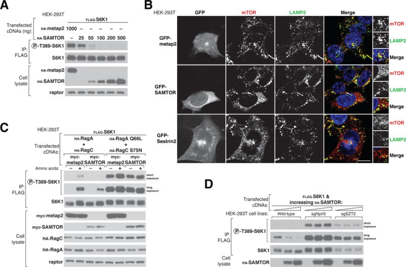
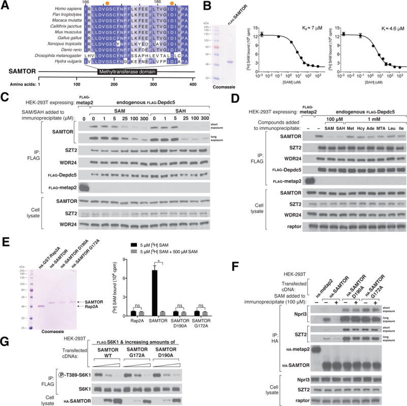
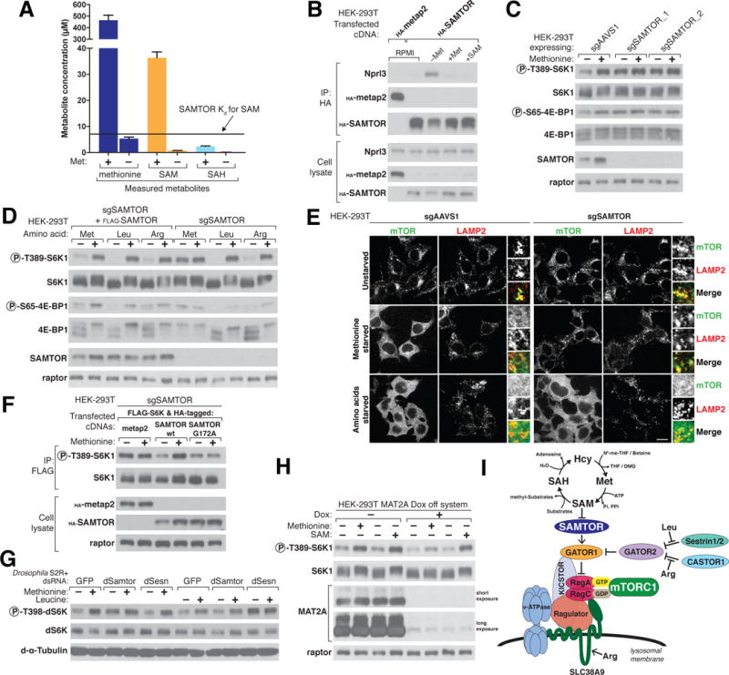
mTOR complex 1 (mTORC1) regulates cell growth and metabolism in response to multiple environmental cues. Nutrients signal via the Rag guanosine triphosphatases (GTPases) to promote the localization of mTORC1 to the lysosomal surface, its site of activation. We identified SAMTOR, a previously uncharacterized protein, which inhibits mTORC1 signaling by interacting with GATOR1, the GTPase activating protein (GAP) for RagA/B. We found that the methyl donor S-adenosylmethionine (SAM) disrupts the SAMTOR-GATOR1 complex by binding directly to SAMTOR with a dissociation constant of approximately 7 μM. In cells, methionine starvation reduces SAM levels below this dissociation constant and promotes the association of SAMTOR with GATOR1, thereby inhibiting mTORC1 signaling in a SAMTOR-dependent fashion. Methionine-induced activation of mTORC1 requires the SAM binding capacity of SAMTOR. Thus, SAMTOR is a SAM sensor that links methionine and one-carbon metabolism to mTORC1 signaling.


Figures

Figure 1
Figure 1. SAMTOR interacts with GATOR1 and KICSTOR
(A) GATOR1 and KICSTOR, but not GATOR2, coimmunoprecipitate SAMTOR. FLAG immunoprecipitates (IP) were prepared from HEK-293T cell lines that stably expressed FLAG-tagged metap2 or Kaptin, or had endogenously FLAG-tagged Depdc5 or WDR59. FLAG immunoprecipitates and lysates were analyzed by immunoblotting for the indicated proteins. FLAG-metap2 served as a negative control. Depdc5 and Nprl3, WDR59 and WDR24, and Kaptin and SZT2 were used as representative components of the GATOR1, GATOR2, and KICSTOR complexes, respectively; Raptor was used as a loading control. Short or long exposure indicates relative blot exposure times. (B) SAMTOR coimmunoprecipitates GATOR1 and KICSTOR, and the interaction requires both GATOR1 and KICSTOR but not GATOR2. FLAG immunoprecipitates were prepared from wild-type, Nprl3-deficient, SZT2- deficient, or WDR24-deficient HEK-293T cells transiently expressing the indicated cDNAs. FLAG immunoprecipitates and lysates were analyzed as in (A). (C) Model showing how SAMTOR interacts with GATOR1 and KICSTOR. (D) Presence or absence of gene orthologs of SAMTOR in several model organisms.
Figure 2
Figure 2. SAMTOR is a negative regulator of mTORC1 signaling that acts upstream of the Rag GTPases, GATOR1, and KICSTOR
(A) Transient overexpression of SAMTOR inhibits mTORC1 signaling. FLAG immunoprecipitates were prepared from HEK-293T cells transfected with 2 ng of FLAG-S6K1 cDNA along with either hemagglutinin (HA)–tagged metap2 cDNA or increasing amounts of HA-SAMTOR cDNA. FLAG immunoprecipitates and cell lysates were analyzed by immunoblotting for the phosphorylation states and levels of the indicated proteins. (B) Overexpression of GFP-SAMTOR displaces mTOR from lysosomes, similar to the effect of GFP-Sestrin2.Wild-type HEK-293Tcells transiently expressing GFP-metap2, GFP-SAMTOR, or GFP-Sestrin2 were processed for immunofluorescence detection of mTOR and the lysosomal marker LAMP2. In all images, insets represent selected fields magnified 5.12X as well as their overlays. Scale bar, 10 μm. (C) SAMTOR functions upstreamof the Rag GTPases to regulate the mTORC1 pathway. HEK-293Tcells expressing the indicated cDNAs were starved of amino acids for 50 min or starved and restimulated with amino acids for 10 min. FLAG immunoprecipitates and cell lysates were analyzed as in (A). (D) SAMTOR functions upstream of GATOR1 and KICSTOR. FLAG immunoprecipitates and cell lysates prepared from wild-type, Nprl3-deficient, or SZT2-deficient HEK-293Tcell lines expressing the indicated cDNAs were analyzed as in (A).
Figure 3
Figure 3. S-adenosylmethionine binds SAMTOR to disrupt its interaction with GATOR1 and KICSTOR
(A) Schematic of the human SAMTOR protein indicating the class I Rossmann fold methyltransferase domain. Shown is an alignment of partial sequences of this domain from SAMTOR in indicated species. Amino acid positions are colored from white to blue in order of increasing sequence similarity. Orange dots denote the Gly172 and Asp190 residues of human SAMTOR. (B) SAMTOR binds SAM and SAH. Purified FLAG-SAMTOR protein was analyzed by SDS–polyacrylamide gel electrophoresis followed by Coomassie blue staining. Binding assays were performed with purified FLAG-SAMTOR incubated with the indicated concentrations of [3H]SAM, unlabeled SAM, or SAH.Values for each point are means ± SD of three technical replicates fromone representative experiment. The experiment was performed twice. (C) SAM and SAH disrupt the interaction of SAMTOR with GATOR1 in vitro. FLAG immunoprecipitates were prepared from endogenously FLAG-tagged Depdc5 HEK-293Tcells. SAM and SAH were added directly to the immunoprecipitates at the indicated concentrations. FLAG immunoprecipitates and cell lysates were analyzed by immunoblotting for the levels of the indicated proteins. (D) The interaction between SAMTOR and GATOR1 is disrupted by 100 μM SAM or SAH, but not by 1mM methionine, homocysteine, adenosine, 5-methylthioadenosine, leucine, or isoleucine. The experiment was performed and analyzed as in (C). (E) Wild-type HA-SAMTOR, but not HA-SAMTOR G172A or D190A, binds SAM. HA-tagged wild-type and mutant SAMTOR proteins were prepared from HEK-293Tcells expressing the indicated cDNAs, and binding assays were performed as in (B). A representative experiment is shown; values are means ± SD of three technical replicates.Two-tailed t tests were used for comparisons between two groups. *P < 0.001; ns, not significant. The experiment was repeated three times. (F) HA-SAMTOR G172A and D190A coimmunoprecipitate more endogenous GATOR1 and KICSTOR than does wild-type SAMTOR, and the interactions are insensitive to SAM added in vitro. HA immunoprecipitates and cell lysates were prepared from HEK-293Tcells transiently expressing wild-type HA-SAMTOR or its mutants G172A or D190A. SAM was added to the immunoprecipitates where indicated. HA immunoprecipitates and cell lysates were analyzed as in (C). (G) HA-SAMTOR G172A and D190A inhibit mTORC1 activity to similar extents as wild-type SAMTOR. FLAG immunoprecipitates were prepared from HEK-293Tcells transfected with the indicated cDNAs. FLAG immunoprecipitates and cell lysates were analyzed by immunoblotting for the phosphorylation states and levels of the indicated proteins.
Figure 4
Figure 4. SAMTOR senses SAM to signal methionine sufficiency to mTORC1
(A) HEK-293Tcells were incubated with or withoutmethionine for 2 hours before sample preparation for liquid chromatography/mass spectrometry (LC/MS)– based measurements of the absolute amounts of the indicated metabolites. The dissociation constant Kd of SAMTOR for SAM is indicated. (B) Methionine starvation increases the interaction between SAMTOR and GATOR1. HEK-293T cells transiently expressing HA-tagged metap2 or SAMTOR were kept in growth medium (RPMI) or starved of methionine for 2 hours (–Met) and then restimulated for 20 min with 100 μM methionine (+Met) or 1 mM SAM (+SAM). HA immunoprecipitates and cell lysates were analyzed by immunoblotting for the levels of the indicated proteins. (C) In SAMTOR-depleted cells, the mTORC1 pathway is resistant to methionine starvation. HEK-293Tcells stably coexpressing Cas9 and the indicated guides were incubated in media with or without methionine for 2 hours. Cell lysates were analyzed by immunoblotting for the phosphorylation states and the levels of the indicated proteins. (D) The loss of SAMTOR does not affect the sensitivity of the mTORC1 pathway to leucine or arginine starvation. SAMTOR-deficient HEK-293Tcells with or without FLAG-SAMTOR expression were starved of the indicated amino acid for 2 hours. Cell lysates were analyzed as in (C). (E) In cells without SAMTOR, mTOR colocalizes with lysosomes even upon methionine starvation. SAMTOR deficient or control HEK-293Tcells were treated as indicated for 2 hours before marker LAMP2. In all images, insets represent selected fields magnified 3.07X as well as their overlays. Scale bar, 10 μm. (F) Reexpression in SAMTOR-null cells of wild-type SAMTOR, but not the SAM-binding G172A mutant of SAMTOR, restored the capacity of the mTORC1 pathway to sense methionine sufficiency. SAMTOR-null cells were transfected with the indicated cDNAs and the cells were treated as in (C) before preparing lysates and FLAG immunoprecipitates. FLAG immunoprecipitates and cell lysates were analyzed as in (C). (G) In Drosophila S2R+ cells depleted of dSamtor or dSesn, the dTOR pathway is resistant to methionine or leucine starvation, respectively. S2R+ cells were transfected with dsRNAs targeting the indicated mRNAs and starved of the indicated amino acids for 1 hour. Cell lysates were analyzed as in (C). (H) Acute loss of MAT2A using a doxycycline-suppressible (dox-off) system attenuates the capacity of mTORC1 to sense methionine but leaves SAM signaling largely intact. MAT2A dox-off HEK-293Tcells were treated with doxycycline (30 ng/ml) for 50 hours before starving them as in (C). Cell lysates were analyzed as in (C). (I) Model depicting how SAM sensing by SAMTOR signals methionine levels to mTORC1. Substrates receiving a methyl group from SAM include DNA, RNA, proteins, and phospholipids. N5-me-THF, N5- methyl-tetrahydrofolate; DMG, dimethylglycine; Pi, inorganic phosphate; PPi, pyrophosphate.

References

    1. Saxton RA, Sabatini DM. mTOR Signaling in Growth, Metabolism, and Disease. Cell. 2017;168:960–976. - PMC - PubMed
    1. Dibble CC, Manning BD. Signal integration by mTORC1 coordinates nutrient input with biosynthetic output. Nat Cell Biol. 2013;15:555–564. - PMC - PubMed
    1. Jewell JL, Russell RC, Guan KL. Amino acid signalling upstream of mTOR. Nature reviews Molecular cell biology. 2013;14:133–139. - PMC - PubMed
    1. Sancak Y, et al. The Rag GTPases bind raptor and mediate amino acid signaling to mTORC1. Science. 2008;320:1496–1501. - PMC - PubMed
    1. Kim E, Goraksha-Hicks P, Li L, Neufeld TP, Guan KL. Regulation of TORC1 by Rag GTPases in nutrient response. Nat Cell Biol. 2008;10:935–945. - PMC - PubMed

Publication types

MeSH terms

Substances