. 2017 Oct 24:11:3045-3063.
doi: 10.2147/DDDT.S144415. eCollection 2017.

10 H-3,6-Diazaphenothiazine induces G2/M phase cell cycle arrest and caspase-dependent apoptosis and inhibits cell invasion of A2780 ovarian carcinoma cells through the regulation of NF-κB and (BIRC6-XIAP) complexes

Affiliations

10 H-3,6-Diazaphenothiazine induces G2/M phase cell cycle arrest and caspase-dependent apoptosis and inhibits cell invasion of A2780 ovarian carcinoma cells through the regulation of NF-κB and (BIRC6-XIAP) complexes

Jianxin Zhang et al. Drug Des Devel Ther. .

Abstract

The asymptomatic properties and high treatment resistance of ovarian cancer result in poor treatment outcomes and high mortality rates. Although the fundamental chemotherapy provides promising anticancer activities, it is associated with severe side effects. The derivative of phenothiazine, namely, 10H-3,6-diazaphenothiazine (PTZ), was synthesized and reported with ideal anticancer effects in a previous paper. In this study, detailed anticancer properties of PTZ was examined on A2780 ovarian cancer cells by investigating the cytotoxicity profiles, mechanism of apoptosis, and cell invasion. Research outcomes revealed PTZ-induced dose-dependent inhibition on A2780 cancer cells (IC50 =0.62 µM), with significant less cytotoxicity toward HEK293 normal kidney cells and H9C2 normal heart cells. Generation of reactive oxygen species (ROS) and polarization of mitochondrial membrane potential (ΔΨm) suggests PTZ-induced cell death through oxidative damage. The RT2 Profiler PCR Array on apoptosis pathway demonstrated PTZ-induced apoptosis via intrinsic (mitochondria-dependent) and extrinsic (cell death receptor-dependent) pathway. Inhibition of NF-κB and subsequent inhibition of (BIRC6-XIAP) complex activities reduced the invasion rate of A2780 cancer cells penetrating through the Matrigel™ Invasion Chamber. Lastly, the cell cycle analysis hypothesizes that the compound is cytostatic and significantly arrests cell proliferation at G2/M phase. Hence, the exploration of the underlying anticancer mechanism of PTZ suggested its usage as promising chemotherapeutic agent.

Keywords: anticancer; cancer cell invasion; mitochondrial function disruption; ovarian cancer; oxidative damage; programmed cell death.


Conflict of interest statement

Disclosure The authors report no conflicts of interest in this work.

Figures

Figure 1
Figure 1
The chemical structure of 10H-3,6-diazaphenothiazine. ©2016 Informa UK Limited, trading as Taylor & Francis Group. Reproduced from Morak-Młodawska B, Pluta K, Latocha M, Suwińka K, Jeleń M, Kuśmierz D. 3,6-Diazaphenothiazines as potential lead molecules – synthesis, characterization and anticancer activity. J Enzyme Inhib Med Chem. 2016;31:1512–1519.
Figure 2
Figure 2
The trial compound PTZ induced concentration-dependent inhibitory effects toward A2780 ovarian cancer cells after incubated for 24 hours. Notes: The results are expressed as mean percentage of living cells over untreated control, and the error bars indicate standard deviation. ***Each treated concentration is significantly different toward untreated control (0 µM) at p<0.05. Abbreviation: PTZ, 10H-3,6-diazaphenothiazine.
Figure 3
Figure 3
Cell cytotoxicity activity of PTZ toward representative normal cell lines. Notes: PTZ expressed lesser cytotoxicity toward (A) HEK293 human embryonic kidney epithelial cells and (B) H9C2 rat embryonic heart myoblast cells by employing the same protocol as A2780 ovarian cancer cells, thus indicating PTZ is more potent and selective toward cancer cells. The results are expressed as mean percentage of living cells over untreated control, and the error bars indicate standard deviation. ***Each treated concentration is significantly different toward untreated control (0 µM) at p<0.05. Abbreviation: PTZ, 10H-3,6-diazaphenothiazine.
Figure 4
Figure 4
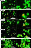
Images of AO/PI staining of A2780 cells. Notes: (A) Untreated, (B) cisplatin-treated, (C) PTZ at 0.3 µM, (D) IC50 of PTZ at 0.62 µM, and (E) PTZ at 0.9 µM; [Left] observation under magnification 200×; [Right] observation under magnification 400× under the same microscopic field. Generally, incubation of PTZ onto A2780 cancer cells for 24 hours gives rise to several features of apoptosis such as fragmentation of DNA and nucleus, condensed chromatin, membrane blebbing, and shrinkage of size of cells. Abbreviations: AO, Acridine orange; PI, propidium iodide; PTZ, 10H-3,6-diazaphenothiazine.
Figure 5
Figure 5
Images of DAPI staining of A2780 ovarian cancer cells. Notes: (A) Untreated, (B) cisplatin-treated, (C) PTZ at 0.3 µM, (D) IC50 of PTZ at 0.62 µM, and (E) PTZ at 0.9 µM; [Left] observation under magnification 200×; [Right] observation under magnification 400× under the same microscopic field. The blue fluorescence intensity was produced upon intercalation of DAPI stain onto DNA fragments. Note that there are less DNA fragments observed in untreated group, in comparison with higher fluorescence intensity (more DNA fragments) observed in treated groups. Hence it is suggested that PTZ initiates apoptosis via induction of DNA damage. Abbreviations: PTZ, 10H-3,6-diazaphenothiazine; DAPI, 4′,6-diamidine-2′-phenylindole.
Figure 6
Figure 6
(A) Untreated, (B) cisplatin-treated, (C) PTZ at 0.3 µM, (D) IC50 of PTZ at 0.62 µM, and (E) PTZ at 0.9 µM. The quadrant graph of quantitative assay of apoptosis induced by PTZ toward A2780 cancer cells. Notes: The quadrant graph of Annexin V-FITC assay is a useful tool to study apoptosis quantitatively. Upon application of PTZ, the percentage of living cells drops drastically and gives a significant increase on the population of apoptotic cells (mainly late apoptosis). Abbreviations: FITC, fluorescein isothiocyanate; PTZ, 10H-3,6-diazaphenothiazine.
Figure 7
Figure 7
Application of PTZ toward A2780 cancer cells results in a significant increase of cellular ROS level (particularly H2O2) up to 129.54% in comparison with the untreated group (by normalized to 100%). Notes: Results are provided as mean from three independent experiments (n=3). ***Indicates results were significantly different to untreated control (0 µM) at p<0.05. Abbreviations: PTZ, 10H-3,6-diazaphenothiazine; ROS, reactive oxygen species.
Figure 8
Figure 8
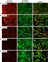
Mitochondrial membrane potential (ΔΨm) of A2780 cancer cells after treated with PTZ was accessed via JC-1 staining assay, with (A) untreated control, (B) cisplatin, (C) 0.30 µM, (D) 0.62 µM, and (E) 0.90 µM. Notes: Noted in untreated group, the high red fluorescence intensity indicated living cells with high mitochondria activity with minimal ΔΨm polarization. The red fluorescence was produced upon binding of JC-1 dye onto non-polarized mitochondrial membrane. Application of PTZ onto A2780 cancer cells resulting polarization of ΔΨm, thus reducing probability of JC-1 bound onto mitochondrial membrane thus generating green fluorescence. Reduction on red/green fluorescence ratio from (C) to (E) are results from polarization of ΔΨm, suggesting PTZ induced growth inhibition toward A2780 cancer cells via disruption on ΔΨm and consequently induced oxidative stress and damages. Magnification: 200×. Abbreviation: PTZ, 10H-3,6-diazaphenothiazine.
Figure 9
Figure 9
Graphical data representing measurement of (A) caspase-3/7, (B) caspase-8, and (C) caspase-9 activities (in percentage) of A2780 cells after induction of apoptosis by PTZ at three reference concentrations. Notes: Results are illustrated as mean from three independent experiments (n=3). ***Indicates results were significantly different to untreated control (0 µM) at p<0.05. Abbreviations: FITC, fluorescein isothiocyanate; PTZ, 10H-3,6-diazaphenothiazine.
Figure 10
Figure 10
The histogram representing cell cycle phase analysis on A2780 cells after treated by PTZ for 24 hours. Notes: (A) Untreated, (B) cisplatin-treated, (C) PTZ at 0.3 µM, (D) IC50 of PTZ at 0.62 µM, and (E) PTZ at 0.9 µM. The trail compound PTZ inhibited proliferation of A2780 cells via induction of G2/M-phase cell cycle checkpoint. Note that the increase of concentration of PTZ results in transition of cell population from G0/G1 phase and lastly accumulated in G2/M phase. Abbreviation: PTZ, 10H-3,6-diazaphenothiazine.
Figure 11
Figure 11
(A) Microscopic images of invaded A2780 cancer cells through the Matrigel™ invasion chamber. By normalizing the invasion rate of cancer cells in untreated control as 100%, application of cisplatin and PTZ exhibited suppression of cancer cells invasion. Photos were taken at a magnification of 200× and each image are representative of three independent experiments. (B) Graphical representation of percentage of invaded cells per microscopic views. The number of invaded cells were calculated by using the cell counter from five random microscopic field under the same microscopic view. ***Indicates results were significantly different to untreated control (0 µM) at p<0.05. Abbreviation: PTZ, 10H-3,6-diazaphenothiazine.
Figure 12
Figure 12
Proposed apoptosis signaling pathways induced by PTZ in A2780 ovarian cancer cells, based on the findings in the RT2 Profiler PCR array (apoptosis pathway). Notes: Note that the compound induced both intrinsic (mitochondria-dependent) and extrinsic (cell death receptor-dependent) apoptosis pathways, with the generation of oxidative damage in the former pathway. The full list of genes and its respective description are referable at https://www.qiagen.com/us/shop/pcr/primer-sets/rt2-profiler-pcr-arrays/?catno=PAHS-012Z#geneglobe. Abbreviations: CDK, cyclin-dependent kinase; DFFA, DNA fragmentation factor subunit alpha; PTZ, 10H-3,6-diazaphenothiazine; ROS, reactive oxygen species; TNF, tumor necrosis factor; TNFR, TNF receptor; FADD, Fas-associated death domain; TRADD, TNF-associated death domain.

Similar articles

Cited by

References

    1. Torre L, Siegel R, Jemal A. Global Cancer Facts and Figures. 3rd ed. Atlanta, GA, USA: American Cancer Society; 2015.
    1. Lee S, Whang I, Wan Q, et al. Profiles of teleost DNA fragmentation factor alpha and beta from rock bream (Oplegnathus fasciatus): molecular characterization and genomic structure and gene expression in immune stress. Genes Genom. 2016;38:193–204.
    1. Baghbani F, Moztarzadeh F. Bypassing multidrug resistant ovarian cancer using ultrasound responsive doxorubicin/curcumin co-deliver alginate nanodroplets. Col Sur B Biointerfaces. 2017;153:132–140. - PubMed
    1. Rzepecka IK, Szafron LM, Stys A, et al. Prognosis of patients with BRCA1-associated ovarian carcinomas depends on TP53 accumulation status in tumor cells. Gynecol Oncol. 2017;144:369–376. - PubMed
    1. Rossing MA, Wicklund KG, Cushing-Haugen KL, et al. Predictive value of symptoms for early detection of ovarian cancer. J Natl Cancer Inst. 2010;102:222–229. - PMC - PubMed

MeSH terms