

High-dose dexamethasone induced LPS-stimulated rat alveolar macrophages apoptosis
- PMID: 29123381
- PMCID: PMC5661847
- DOI: 10.2147/DDDT.S147014
High-dose dexamethasone induced LPS-stimulated rat alveolar macrophages apoptosis
Abstract
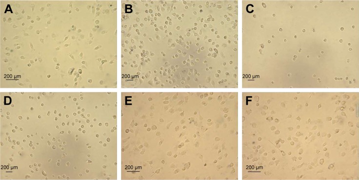
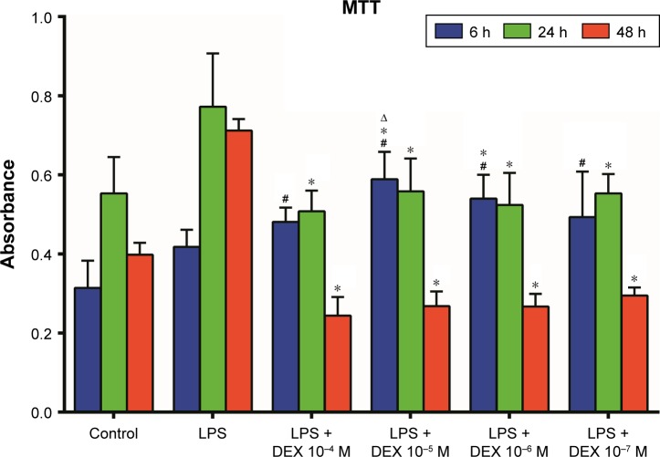
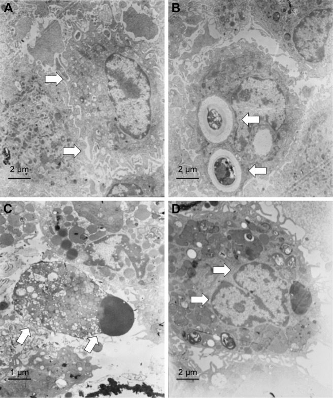
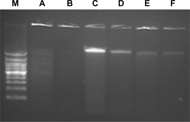
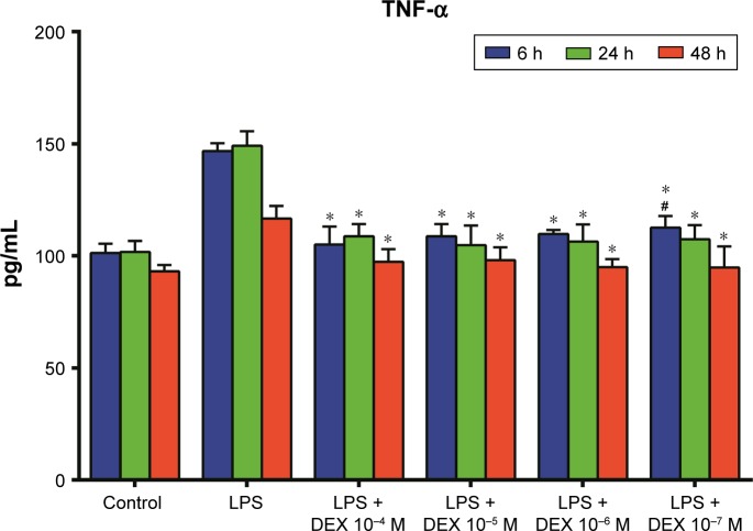
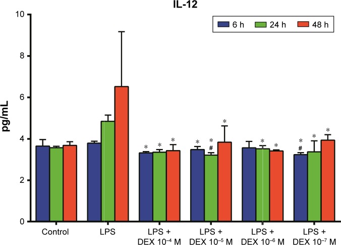
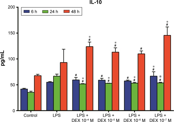
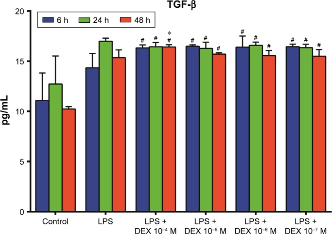
Prolonged administration of an excessive dose of corticosteroids proved to be harmful for patients with acute lung injury (ALI). A previous study has found that repeated administration of an excessive dose of methylprednisolone reduced alveolar macrophages (AMs) in bronchoalveolar lavage fluid (BALF) with an unknown mechanism. This study aimed to investigate the effect of excessive use of dexamethasone (Dex) on BALF AMs in vitro. Transmission electron microscopy and DNA fragmentation analysis demonstrated that 10-4 and 10-5 M Dex induced lipopolysaccharide-stimulated rat AMs apoptosis with downregulation of tumor necrosis factor-α, interleukin (IL)-12 and upregulation of IL-10, transforming growth factor-β. These results indicated that apoptosis might be a novel contribution involved in the detrimental effect of excessive dose of Dex clinically used to treat ALI.
Keywords: acute lung injury; alveolar macrophage; apoptosis; dexamethasone; inflammatory cytokines; lipopolysaccharide.
Conflict of interest statement
Disclosure The authors report no conflicts of interest in this work.
Figures
Similar articles
-
The harmful effect of prolonged high-dose methylprednisolone in acute lung injury.Int Immunopharmacol. 2013 Feb;15(2):223-6. doi: 10.1016/j.intimp.2012.12.004. Epub 2012 Dec 19. Int Immunopharmacol. 2013. PMID: 23260416
-
Low-dose dexamethasone alleviates lipopolysaccharide-induced acute lung injury in rats and upregulates pulmonary glucocorticoid receptors.Respirology. 2008 Nov;13(6):772-80. doi: 10.1111/j.1440-1843.2008.01344.x. Epub 2008 Jul 24. Respirology. 2008. PMID: 18657064
-
Classically Activated Macrophages Protect against Lipopolysaccharide-induced Acute Lung Injury by Expressing Amphiregulin in Mice.Anesthesiology. 2016 May;124(5):1086-99. doi: 10.1097/ALN.0000000000001026. Anesthesiology. 2016. PMID: 26808632
-
Glucocorticoid combined with hyaluronic acid enhance glucocorticoid receptor activity through inhibiting p-38MAPK signal pathway activation in treating acute lung injury in rats.Eur Rev Med Pharmacol Sci. 2016 Sep;20(18):3920-3929. Eur Rev Med Pharmacol Sci. 2016. PMID: 27735022
-
Effect of in vitro and in vivo administration of dexamethasone on rat macrophage functions: comparison between alveolar and peritoneal macrophages.Eur Respir J. 1996 Feb;9(2):301-6. doi: 10.1183/09031936.96.09020301. Eur Respir J. 1996. PMID: 8777968
Cited by
-
Dexamethasone-induced impairment of post-injury skeletal muscle regeneration.BMC Vet Res. 2019 Feb 11;15(1):56. doi: 10.1186/s12917-019-1804-1. BMC Vet Res. 2019. PMID: 30744624 Free PMC article.
-
Comprehensive study of dexamethasone on albumin biogenesis during normal and pathological renal conditions.Pharm Biol. 2020 Dec;58(1):1252-1262. doi: 10.1080/13880209.2020.1855214. Pharm Biol. 2020. PMID: 33332210 Free PMC article.
-
Dual-target corticosteroid therapy for refractory Ménière's disease: influence of vestibular aqueduct patency and endotypes.Eur Arch Otorhinolaryngol. 2025 Jul 4. doi: 10.1007/s00405-025-09542-2. Online ahead of print. Eur Arch Otorhinolaryngol. 2025. PMID: 40615707 No abstract available.
-
Intranasal Flunisolide Suppresses Pathological Alterations Caused by Silica Particles in the Lungs of Mice.Front Endocrinol (Lausanne). 2020 Jun 17;11:388. doi: 10.3389/fendo.2020.00388. eCollection 2020. Front Endocrinol (Lausanne). 2020. PMID: 32625168 Free PMC article.
-
More Than Suppression: Glucocorticoid Action on Monocytes and Macrophages.Front Immunol. 2019 Aug 27;10:2028. doi: 10.3389/fimmu.2019.02028. eCollection 2019. Front Immunol. 2019. PMID: 31507614 Free PMC article. Review.
References
-
- Zambon M, Vincent JL. Mortality rates for patients with acute lung injury/ARDS have decreased over time. Chest. 2008;133:1120–1127. - PubMed
-
- Bernard GR, Luce JM, Sprung CL, et al. High-dose corticosteroids in patients with the adult respiratory distress syndrome. N Engl J Med. 1987;317:1565–1570. - PubMed
-
- Meduri GU, Headley AS, Golden E, et al. Effect of prolonged methylprednisolone therapy in unresolving acute respiratory distress syndrome: a randomized controlled trial. JAMA. 1998;280:159–165. - PubMed
-
- Steinberg KP, Hudson LD, Goodman RB, et al. Efficacy and safety of corticosteroids for persistent acute respiratory distress syndrome. N Engl J Med. 2006;354:1671–1684. - PubMed
-
- Lohmann-Matthes ML, Steinmüller C, Franke-Ullmann G. Pulmonary macrophages. Eur Respir J. 1994;7:1678–1689. - PubMed
MeSH terms
Substances
LinkOut - more resources
Full Text Sources
Other Literature Sources
Medical
