In vivo analysis of the Escherichia coli ultrastructure by small-angle scattering
- PMID: 29123677
- PMCID: PMC5668860
- DOI: 10.1107/S2052252517013008
In vivo analysis of the Escherichia coli ultrastructure by small-angle scattering
Abstract
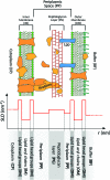
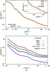
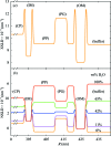
The flagellated Gram-negative bacterium Escherichia coli is one of the most studied microorganisms. Despite extensive studies as a model prokaryotic cell, the ultrastructure of the cell envelope at the nanometre scale has not been fully elucidated. Here, a detailed structural analysis of the bacterium using a combination of small-angle X-ray and neutron scattering (SAXS and SANS, respectively) and ultra-SAXS (USAXS) methods is presented. A multiscale structural model has been derived by incorporating well established concepts in soft-matter science such as a core-shell colloid for the cell body, a multilayer membrane for the cell wall and self-avoiding polymer chains for the flagella. The structure of the cell envelope was resolved by constraining the model by five different contrasts from SAXS, and SANS at three contrast match points and full contrast. This allowed the determination of the membrane electron-density profile and the inter-membrane distances on a quantitative scale. The combination of USAXS and SAXS covers size scales from micrometres down to nanometres, enabling the structural elucidation of cells from the overall geometry down to organelles, thereby providing a powerful method for a non-invasive investigation of the ultrastructure. This approach may be applied for probing in vivo the effect of detergents, antibiotics and antimicrobial peptides on the bacterial cell wall.
Keywords: Escherichia coli; in vivo analysis; small-angle scattering; ultrastructure.
Figures


Similar articles
-
Evolution of the analytical scattering model of live Escherichia coli.J Appl Crystallogr. 2021 Mar 3;54(Pt 2):473-485. doi: 10.1107/S1600576721000169. eCollection 2021 Apr 1. J Appl Crystallogr. 2021. PMID: 33953653 Free PMC article.
-
Use of small-angle X-ray scattering to resolve intracellular structure changes of Escherichia coli cells induced by antibiotic treatment.J Appl Crystallogr. 2016 Dec 1;49(Pt 6):2210-2216. doi: 10.1107/S1600576716018562. eCollection 2016 Dec 1. J Appl Crystallogr. 2016. PMID: 27980516 Free PMC article.
-
Ultra-/Small Angle X-ray Scattering (USAXS/SAXS) and Static Light Scattering (SLS) Modeling as a Tool to Determine Structural Changes and Effect on Growth in S. epidermidis.ACS Appl Bio Mater. 2022 Aug 15;5(8):3703-3712. doi: 10.1021/acsabm.2c00218. Epub 2022 Jul 29. ACS Appl Bio Mater. 2022. PMID: 35905477 Free PMC article.
-
Small-angle scattering techniques for peptide and peptide hybrid nanostructures and peptide-based biomaterials.Adv Colloid Interface Sci. 2023 Aug;318:102959. doi: 10.1016/j.cis.2023.102959. Epub 2023 Jul 7. Adv Colloid Interface Sci. 2023. PMID: 37473606 Review.
-
Recent advances in small-angle scattering and its expanding impact in structural biology.Structure. 2022 Jan 6;30(1):15-23. doi: 10.1016/j.str.2021.09.008. Epub 2021 Dec 13. Structure. 2022. PMID: 34995477 Review.
Cited by
-
Synchrotron Scattering Methods for Nanomaterials and Soft Matter Research.Materials (Basel). 2020 Feb 6;13(3):752. doi: 10.3390/ma13030752. Materials (Basel). 2020. PMID: 32041363 Free PMC article. Review.
-
A multipurpose instrument for time-resolved ultra-small-angle and coherent X-ray scattering.J Appl Crystallogr. 2018 Oct 11;51(Pt 6):1511-1524. doi: 10.1107/S1600576718012748. eCollection 2018 Dec 1. J Appl Crystallogr. 2018. PMID: 30546286 Free PMC article.
-
LetB Structure Reveals a Tunnel for Lipid Transport across the Bacterial Envelope.Cell. 2020 Apr 30;181(3):653-664.e19. doi: 10.1016/j.cell.2020.03.030. Cell. 2020. PMID: 32359438 Free PMC article.
-
Structure-based screening of binding affinities via small-angle X-ray scattering.IUCrJ. 2020 May 6;7(Pt 4):644-655. doi: 10.1107/S2052252520004169. eCollection 2020 Jul 1. IUCrJ. 2020. PMID: 32695411 Free PMC article.
-
Studying the surfaces of bacteria using neutron scattering: finding new openings for antibiotics.Biochem Soc Trans. 2020 Oct 30;48(5):2139-2149. doi: 10.1042/BST20200320. Biochem Soc Trans. 2020. PMID: 33005925 Free PMC article. Review.
References
-
- Asakura, S., Eguchi, G. & Iino, T. (1964). J. Mol. Biol. 10, 42–56. - PubMed
-
- Calladine, C. R. (1978). J. Mol. Biol. 118, 457–479.
LinkOut - more resources
Full Text Sources
Other Literature Sources

