. 2017 Oct 9;4(Pt 6):769-777.
doi: 10.1107/S2052252517013124. eCollection 2017 Nov 1.

Mix-and-diffuse serial synchrotron crystallography

Affiliations

Mix-and-diffuse serial synchrotron crystallography

Kenneth R Beyerlein et al. IUCrJ. .

Abstract

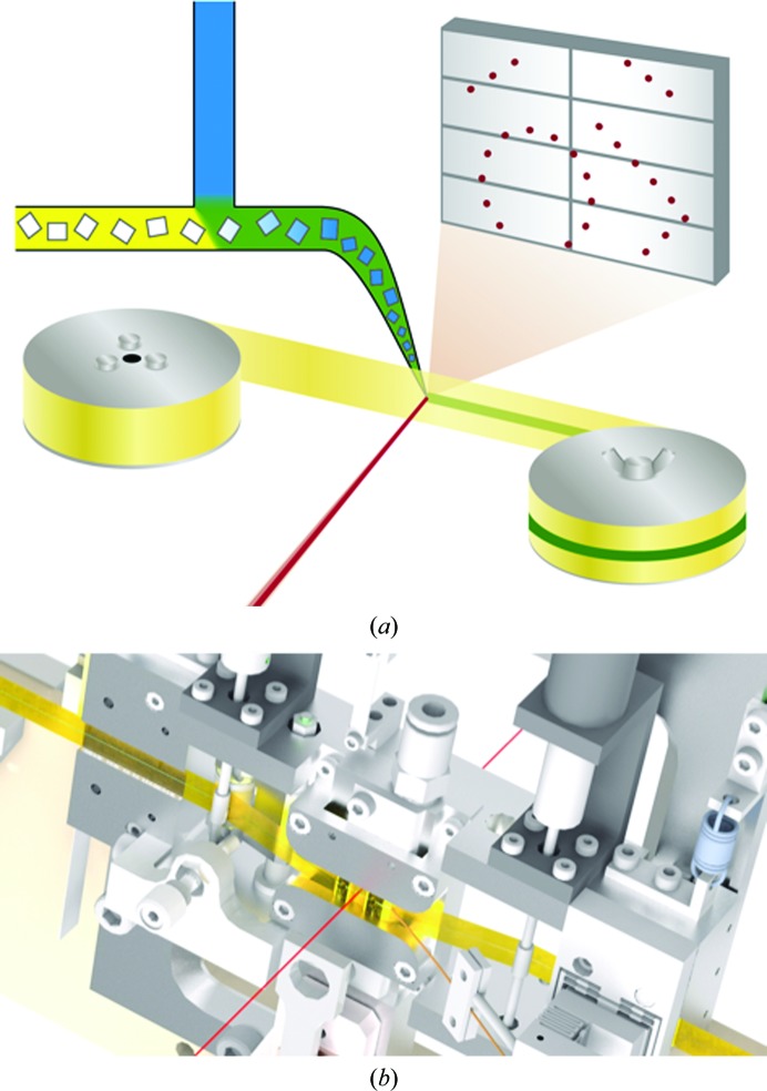
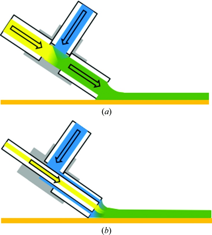
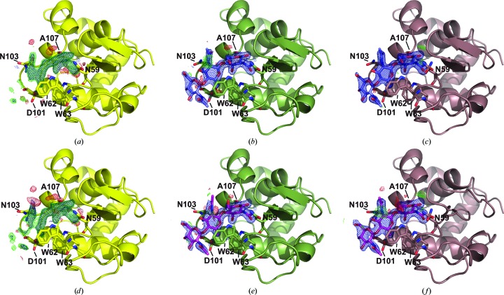
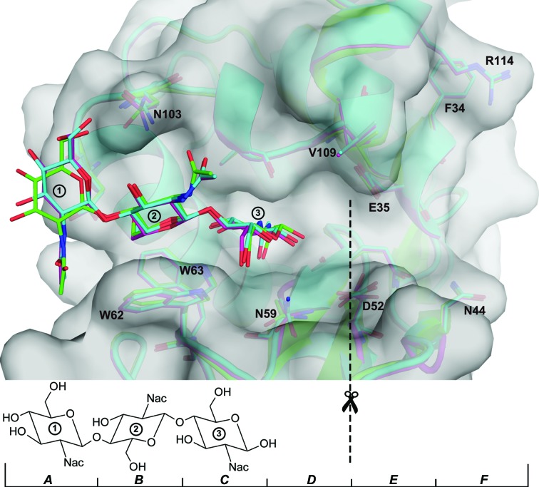
Unravelling the interaction of biological macromolecules with ligands and substrates at high spatial and temporal resolution remains a major challenge in structural biology. The development of serial crystallography methods at X-ray free-electron lasers and subsequently at synchrotron light sources allows new approaches to tackle this challenge. Here, a new polyimide tape drive designed for mix-and-diffuse serial crystallography experiments is reported. The structure of lysozyme bound by the competitive inhibitor chitotriose was determined using this device in combination with microfluidic mixers. The electron densities obtained from mixing times of 2 and 50 s show clear binding of chitotriose to the enzyme at a high level of detail. The success of this approach shows the potential for high-throughput drug screening and even structural enzymology on short timescales at bright synchrotron light sources.

Keywords: X-ray crystallography; drug discovery; lysozyme; protein structure; serial crystallography; time-resolved studies.


Figures

Figure 1
Figure 1
Schematics of the experimental setup. (a) The native protein crystal suspension (yellow) is mixed inline with the substrate solution (blue) before being deposited onto the detector side of the tape. The X-ray beam (red) is focused on the centre of the polyimide tape (yellow ribbon), which is being drawn from the feeder roll on the left to the collector roll on the right. A diffraction pattern measured in transmission is illustrated as red dots on the grey detector. (b) A technical drawing of the tape-drive device shows how tape is held under tension between two rollers near the interaction region and that X-rays pass through a hole in the centre of the device.
Figure 2
Figure 2
The mixer configurations for the (a) 50 s and (b) 2 s mixing times are illustrated, showing a side view of the sample-stream deposition on the tape (orange line). The colour scheme for the respective liquids is the same as in the experimental setup schematic (Fig. 1 ▸ a). The images are not to scale.
Figure 3
Figure 3
CTO bound to lysozyme: short-mixing case (a, b, c) and long-mixing case (d, e, f). (a) and (d) show the 2F oF c (blue, 1σ) and F oF c (green and red, 2.5σ) maps of the lysozyme binding site after initial refinement with ligand-free phases. (b) and (e) show the maps at the same levels after automatic ligand-placement and refinement, and (c) and (f) show the respective final refined models and maps. Residues forming hydrogen bonds to CTO are shown.
Figure 4
Figure 4
Comparison of ligand binding in the long-mixing case solved starting from PDB entries 4et8 (magenta) and 1hew (cyan) with that in the co-crystallized ligand structure PDB entry 1hew (green). It can be seen that all ligands bind to subsites ABC and that only the most flexible sugar ring 1 shows a significantly different orientation. Most notably, the different orientation of the hydroxyl O atom at position 6 of the pyranose ring and of the acetamido group of Asn103 results in a hydrogen bond between the ligand and protein in PDB entry 1hew but not in the mixing cases. Binding-site residues of lysozyme are displayed and the associated binding subsites AF are indicated at the bottom of the figure. The dashed line indicates the active site for catalysis (cleavage site).

Similar articles

Cited by

  • Flow-Xl: a new facility for the analysis of crystallization in flow systems.
    Turner TD, O'Shaughnessy C, He X, Levenstein MA, Hunter L, Wojciechowski J, Bristowe H, Stone R, Wilson CC, Florence A, Robertson K, Kapur N, Meldrum FC. Turner TD, et al. J Appl Crystallogr. 2024 Aug 19;57(Pt 5):1299-1310. doi: 10.1107/S1600576724006113. eCollection 2024 Oct 1. J Appl Crystallogr. 2024. PMID: 39387089 Free PMC article.
  • Serial protein crystallography in an electron microscope.
    Bücker R, Hogan-Lamarre P, Mehrabi P, Schulz EC, Bultema LA, Gevorkov Y, Brehm W, Yefanov O, Oberthür D, Kassier GH, Dwayne Miller RJ. Bücker R, et al. Nat Commun. 2020 Feb 21;11(1):996. doi: 10.1038/s41467-020-14793-0. Nat Commun. 2020. PMID: 32081905 Free PMC article.
  • Enzyme intermediates captured "on the fly" by mix-and-inject serial crystallography.
    Olmos JL Jr, Pandey S, Martin-Garcia JM, Calvey G, Katz A, Knoska J, Kupitz C, Hunter MS, Liang M, Oberthuer D, Yefanov O, Wiedorn M, Heyman M, Holl M, Pande K, Barty A, Miller MD, Stern S, Roy-Chowdhury S, Coe J, Nagaratnam N, Zook J, Verburgt J, Norwood T, Poudyal I, Xu D, Koglin J, Seaberg MH, Zhao Y, Bajt S, Grant T, Mariani V, Nelson G, Subramanian G, Bae E, Fromme R, Fung R, Schwander P, Frank M, White TA, Weierstall U, Zatsepin N, Spence J, Fromme P, Chapman HN, Pollack L, Tremblay L, Ourmazd A, Phillips GN Jr, Schmidt M. Olmos JL Jr, et al. BMC Biol. 2018 May 31;16(1):59. doi: 10.1186/s12915-018-0524-5. BMC Biol. 2018. PMID: 29848358 Free PMC article.
  • Data reduction for serial crystallography using a robust peak finder.
    Hadian-Jazi M, Sadri A, Barty A, Yefanov O, Galchenkova M, Oberthuer D, Komadina D, Brehm W, Kirkwood H, Mills G, de Wijn R, Letrun R, Kloos M, Vakili M, Gelisio L, Darmanin C, Mancuso AP, Chapman HN, Abbey B. Hadian-Jazi M, et al. J Appl Crystallogr. 2021 Sep 13;54(Pt 5):1360-1378. doi: 10.1107/S1600576721007317. eCollection 2021 Oct 1. J Appl Crystallogr. 2021. PMID: 34667447 Free PMC article.
  • Beef tallow injection matrix for serial crystallography.
    Nam KH. Nam KH. Sci Rep. 2022 Jan 13;12(1):694. doi: 10.1038/s41598-021-04714-6. Sci Rep. 2022. PMID: 35027663 Free PMC article.

References

    1. Adams, P. D. et al. (2010). Acta Cryst. D66, 213–221. - PubMed
    1. Afonine, P. V., Grosse-Kunstleve, R. W., Echols, N., Headd, J. J., Moriarty, N. W., Mustyakimov, M., Terwilliger, T. C., Urzhumtsev, A., Zwart, P. H. & Adams, P. D. (2012). Acta Cryst. D68, 352–367. - PMC - PubMed
    1. Barends, T. R. M. et al. (2015). Science, 350, 445–450. - PubMed
    1. Barends, T. R. M., Foucar, L., Botha, S., Doak, R. B., Shoeman, R. L., Nass, K., Koglin, J. E., Williams, G. J., Boutet, S., Messerschmidt, M. & Schlichting, I. (2014). Nature (London), 505, 244–247. - PubMed
    1. Blake, C. C. F., Johnson, L. N., Mair, G. A., North, A. C. T., Phillips, D. C. & Sarma, V. R. (1967). Proc. R. Soc. B Biol. Sci. 167, 378–388. - PubMed