

Lemierre syndrome involving external jugular vein
- PMID: 29123694
- PMCID: PMC5667194
- DOI: 10.1002/ams2.61
Lemierre syndrome involving external jugular vein
Abstract
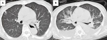
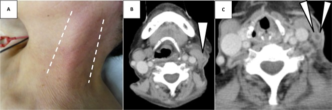
Case: A 74-year-old woman with a week-long history of cold symptoms was diagnosed with Lemierre syndrome that involved her left external jugular vein.
Outcome: The patient was successfully treated with 4 weeks of antibiotics and anticoagulant treatment. Typical cases of Lemierre syndrome involve only the internal jugular vein. The external jugular vein is anatomically distant from the pharyngolaryngeal space and usually does not receive blood or lymphatic flow from there. Thus, Lemierre syndrome ordinarily does not involve the external jugular vein and clinical characteristics of external jugular vein-involving Lemierre syndrome have not been uncovered, mainly due to its rarity. Based on our review, it would not much differ from those of typical cases.
Conclusion: Considering the potential severity and mortality, more attention should be paid to this potentially fatal disease that may demonstrate atypical manifestation, as shown in this case. Accumulation of cases would be needed for further understanding.
Keywords: External jugular vein (EJV); Fusobacterium; Lemierre syndrome; jugular vein suppurative thrombophlebitis; septic pulmonary emboli (SPE).
Figures
Similar articles
-
A Rare Case Report of Lemierre Syndrome from the Anterior Jugular Vein.Clin Pract Cases Emerg Med. 2020 Aug;4(3):454-457. doi: 10.5811/cpcem.2020.7.47442. Clin Pract Cases Emerg Med. 2020. PMID: 32926711 Free PMC article.
-
Atypical Lemierre syndrome.Eur Ann Otorhinolaryngol Head Neck Dis. 2016 Apr;133(2):123-4. doi: 10.1016/j.anorl.2015.12.001. Epub 2015 Dec 21. Eur Ann Otorhinolaryngol Head Neck Dis. 2016. PMID: 26718846
-
[Pleuro-pneumonia revealing septic thrombophlebitis of the jugular vein: Think about the Lemierre's syndrome].Rev Mal Respir. 2016 Jan;33(1):72-7. doi: 10.1016/j.rmr.2015.05.006. Epub 2015 Jul 7. Rev Mal Respir. 2016. PMID: 26163394 French.
-
Lemierre syndrome complicating otitis externa: case report and literature review.J Emerg Med. 2012 Apr;42(4):e77-80. doi: 10.1016/j.jemermed.2009.02.014. Epub 2009 Mar 27. J Emerg Med. 2012. PMID: 19327936 Review.
-
[Diagnostic imaging in Lemierre's+ syndrome].Enferm Infecc Microbiol Clin. 1993 May;11(5):263-6. Enferm Infecc Microbiol Clin. 1993. PMID: 8324024 Review. Spanish.
Cited by
-
Isolated external jugular thrombophlebitis secondary to acute pharyngitis: a case report and a review of the literature.Ital J Pediatr. 2024 Sep 16;50(1):179. doi: 10.1186/s13052-024-01760-4. Ital J Pediatr. 2024. PMID: 39285285 Free PMC article. Review.
-
An unusual case of cavitating pulmonary nodules: Lemierre's syndrome with isolated involvement of the external jugular vein.BJR Case Rep. 2018 Feb 22;4(3):20170093. doi: 10.1259/bjrcr.20170093. eCollection 2018 Mar. BJR Case Rep. 2018. PMID: 31489210 Free PMC article.
-
Management of Patients Receiving Anticoagulation Therapy in Dental Practice: A Systematic Review.Healthcare (Basel). 2024 Aug 2;12(15):1537. doi: 10.3390/healthcare12151537. Healthcare (Basel). 2024. PMID: 39120240 Free PMC article. Review.
-
Pediatric Patient with Lemierre Syndrome of the External Jugular Vein: Case Report and Literature Review.Int Arch Otorhinolaryngol. 2021 Feb 19;25(4):e633-e640. doi: 10.1055/s-0040-1721337. eCollection 2021 Oct. Int Arch Otorhinolaryngol. 2021. PMID: 34737835 Free PMC article.
-
Lemierre's syndrome by Bacillus circulans, Fusobacterium nucleatum and Staphylococcus aureus with involvement of the internal and external jugular vein.Germs. 2021 Jun 2;11(2):314-318. doi: 10.18683/germs.2021.1267. eCollection 2021 Jun. Germs. 2021. PMID: 34422702 Free PMC article.
References
-
- Hagelskjaer LH, Prag J, Malczynski J, Kristensen JH. Incidence and clinical epidemiology of necrobacillosis, including Lemierre's syndrome, in Denmark 1990–95. Eur. J. Clin. Microbiol. Infect. Dis. 1998; 17: 561–565. - PubMed
-
- Stokroos RJ, Manni JJ, de Kruijk JR, Soudijn ER. Lemierre syndrome and acute mastoiditis. Arch. Otolaryngol. Head Neck Surg. 1999; 125: 589–591. - PubMed
-
- Schwartz HC, Nguyen DC. Postanginal septicaemia with external jugular venous thrombosis: case report. Br. J. Oral Maxillofac. Surg. 1999; 37: 144–146. - PubMed
-
- Shibasaki Warabi Y, Yoshikawa H, Idezuka J, Yamazaki M, Onishi Y. Cerebral infarctions and brain abscess due to Lemierre syndrome. Intern. Med. 2005; 44: 653–656. - PubMed
-
- Abe H, Kisara A, Yagishita Y. A case of Lemierre syndrome. ICU & CCU. 1998; 22: 281–285.
Publication types
LinkOut - more resources
Full Text Sources
Other Literature Sources
