

Severe heatstroke complicated with Takotsubo cardiomyopathy
- PMID: 29123776
- PMCID: PMC5667366
- DOI: 10.1002/ams2.151
Severe heatstroke complicated with Takotsubo cardiomyopathy
Abstract
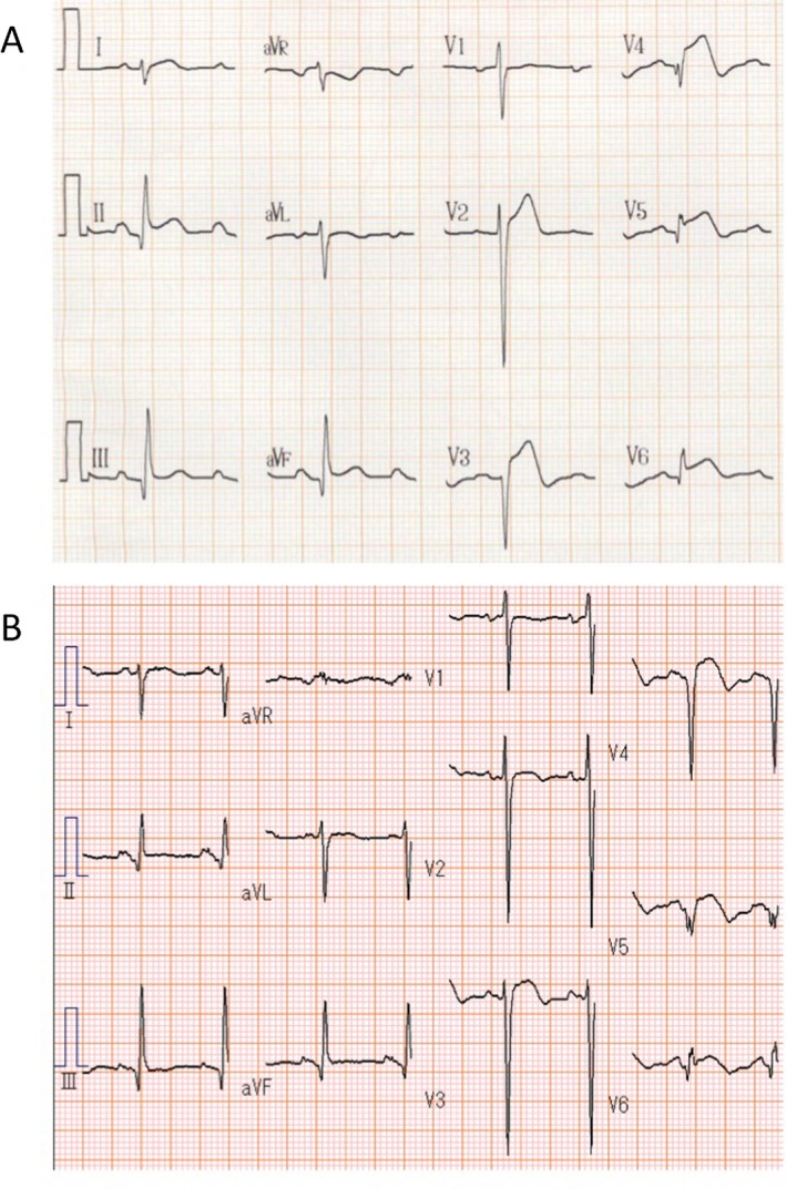
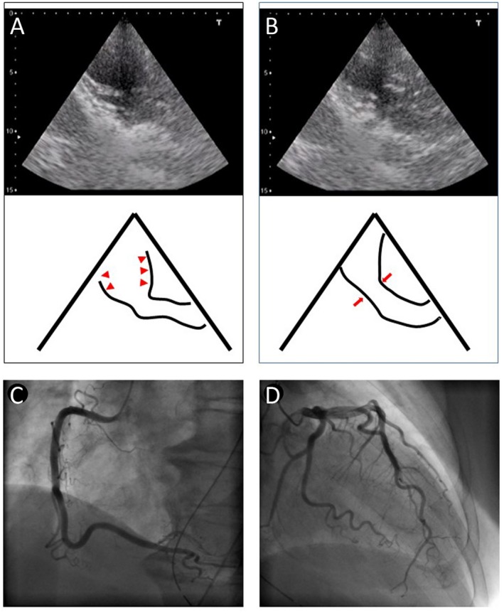
Case: A 69 year-old female with history of schizophrenia was transported to our hospital by ambulance due to coma. On arrival, she was hypotensive and tachycardic with a Glasgow coma scale score of 3 and a rectal core temperature of 40°C. Heatstroke was strongly suspected as the cause of the coma and hypotension. Active external cooling with an electric fan and cooled IV fluid administration were started. Her electrocardiogram (EKG) showed ST elevation in V2-6, II, III and aVF. Echocardiography revealed apical ballooning, which indicated Takotsubo cardiomyopathy. Coronary angiography indicated normal coronary arteries.
Outcome: After admission to the intensive care unit, her cardiovascular status gradually improved and she was transferred to the psychiatric ward on day 36.
Conclusion: Heatstroke and Takotsubo cardiomyopathy can share the same pathophysiology. Close evaluation of hemodynamic status and myocardial damage is critical for survival.
Keywords: Circulation; Takotsubo cardiomyopathy; echocardiography; heatstroke; shock.
Figures
Similar articles
-
Takotsubo cardiomyopathy complicated with apical thrombus formation on first day of the illness: a case report and literature review.BMC Cardiovasc Disord. 2017 Jul 3;17(1):176. doi: 10.1186/s12872-017-0616-0. BMC Cardiovasc Disord. 2017. PMID: 28673245 Free PMC article. Review.
-
Zumba-induced Takotsubo cardiomyopathy: a case report.J Med Case Rep. 2018 Jun 10;12(1):160. doi: 10.1186/s13256-018-1696-x. J Med Case Rep. 2018. PMID: 29885664 Free PMC article.
-
Basal wall hypercontraction of Takotsubo cardiomyopathy in a patient who had been diagnosed with dilated cardiomyopathy: a case report.BMC Cardiovasc Disord. 2017 Dec 12;17(1):293. doi: 10.1186/s12872-017-0730-z. BMC Cardiovasc Disord. 2017. PMID: 29233129 Free PMC article.
-
Takotsubo cardiomyopathy in a Caucasian Italian woman: case report.Cardiovasc Ultrasound. 2007 Apr 6;5:18. doi: 10.1186/1476-7120-5-18. Cardiovasc Ultrasound. 2007. PMID: 17417970 Free PMC article.
-
Takotsubo Cardiomyopathy presenting with different morphological patterns in the same patient: a case report and review of the literature.Cardiovasc Pathol. 2020 Jul-Aug;47:107204. doi: 10.1016/j.carpath.2020.107204. Epub 2020 Jan 15. Cardiovasc Pathol. 2020. PMID: 32169829 Review.
Cited by
-
Acute Myocarditis in a Patient with Exertional Heat Illness: A Rare Association.Eur J Case Rep Intern Med. 2020 Nov 6;7(12):002027. doi: 10.12890/2020_002027. eCollection 2020. Eur J Case Rep Intern Med. 2020. PMID: 33457364 Free PMC article.
-
Multiparametric cardiac magnetic resonance reveals persistent myocardial inflammation in patients with exertional heat illness.Eur Radiol. 2023 Nov;33(11):8165-8176. doi: 10.1007/s00330-023-09706-w. Epub 2023 May 5. Eur Radiol. 2023. PMID: 37145150
References
-
- Yeo TP. Heatstroke a comprehensive review. AACN Clin. Issues 2004; 15: 280–293. - PubMed
-
- Zahger D, Moses A, Weiss AT. Evidence of prolonged myocardial dysfunction in heatstroke. Chest 1989; 95: 1089–1091. - PubMed
-
- Chen W‐T, Lin C‐H, Hsieh M‐H et al Stress‐induced cardiomyopathy caused by heatstroke. Ann. Emerg. Med. 2012; 60: 63–66. - PubMed
-
- Henderson A, Simon JW, Melia WM et al Heat illness. A report of 45 cases from Hong Kong. J. R. Army Med. Corps 1986; 132: 76–84. - PubMed
Publication types
LinkOut - more resources
Full Text Sources
Other Literature Sources
