Review
. 2016 Feb 26;3(3):219-229.
doi: 10.1002/ams2.188. eCollection 2016 Jul.

The retroperitoneal interfascial planes: current overview and future perspectives

Affiliations
Review

The retroperitoneal interfascial planes: current overview and future perspectives

Kazuo Ishikawa et al. Acute Med Surg. .

Abstract

Recently, the concept of interfascial planes has become the prevalent theory among radiologists for understanding the retroperitoneal anatomy, having replaced the classic tricompartmental theory. However, it is a little known fact that the concept remains incomplete and includes embryological errors, which have been revised on the basis of our microscopic study. We believe that the concept not only provides a much clearer understanding of the retroperitoneal anatomy, but it also allows further development for diagnosis and treatment of retroperitoneal injuries and diseases, should it become an accomplished theory. We explain the history and outline of the concept of interfascial planes, correct common misunderstandings about the concept, explain the unconsciously applied therapeutic procedures based on the concept, and present future perspectives of the concept using our published and unpublished data. This knowledge could be essential to acute care physicians and surgeons sometime soon.

Keywords: Embryology; interfascial planes; retroperitoneum; sepsis/multiple organ failure; trauma.


Figures
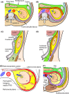
Figure 1
Figure 1
Diagrams of retroperitoneal anatomy. A, Cross‐sectional diagram of the median and left parts of the retroperitoneum according to the tricompartmental theory.3 The retroperitoneum is divided into the anterior pararenal space (green area), perirenal space (yellow area), and posterior pararenal space (gray area) by the anterior renal fascia (red line), posterior renal fascia (purple line), and lateroconal fascia (blue line). Ao, aorta; IVC, inferior vena cava; K, kidney, PM, psoas muscle. B, Cross‐sectional diagram of the same area in panel A, according to the concept of interfascial planes.5 This diagram depicts the interfascial planes as potential spaces among the three compartments: the retromesenteric plane (red area) corresponds to the anterior renal fascia; the retrorenal plane (purple area) corresponds to the posterior renal fascia; and the lateroconal plane (sky‐blue area) corresponds to the lateroconal fascia. Note that the perirenal space is closed medially. C, Longitudinal diagram of the retroperitoneum according to the tricompartmental theory.3 As shown within the dotted circle, the bottom of the perirenal space is patent, and the retroperitoneum below the discontinued renal fasciae (olive area) is ambiguous. D, Longitudinal diagram of the retroperitoneum according to the concept of interfascial planes. The perirenal space is closed inferiorly (open arrow). The combined interfascial plane (pink area) is formed by the inferior blending of the retromesenteric and retrorenal planes and continues into the pelvis. Renal lesions can reach interfascial planes through the perinephric bridging septa (described below) and spread within the combined interfascial plane into the pelvis. E, Aizenstein's advanced diagram of the planes.5 A renal lesion (red area) spreads within the perinephric bridging septa (dotted arrows) and intrudes into the interfascial planes. The lesion can extend contralaterally (red dashed arrow) by way of the retromesenteric plane. F, Our modification of the concept.10, 11 We pointed out that the retrorenal plane, not the posterior pararenal space, lies immediately adjacent to the psoas muscle and quadratus lumborum muscle (QLM) and that the inner border of the posterior pararenal space is limited to the lateral edge of the QLM.10 In addition, we advocated another potential space communicating with the retrorenal plane, which exists behind the posterior pararenal space, and named this the subfascial plane (gold area).10, 11 Panels in this figure are reprinted with permission from the copyright owners (panels A, C: Springer Science+Business Media;3 panels B, E: American Roentgen Ray Society5; panel F: Elsevier11; panel D: Nagai Shoten Co.8, 9 and Wolters Kluwer Health, Inc.10; illustrated by Ishikawa, according to the description by Aizenstein et al.5).
Figure 2
Figure 2
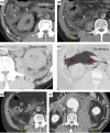
Traumatic retroperitoneal hematoma. A, A 74‐year‐old man with left renal injury. Computed tomography (CT) image on admission shows hematoma sequentially spreading from perinephric bridging septa (thin dotted white arrows) in the perirenal space (Type II) to the retromesenteric (white thick arrows), retrorenal (black triangles), and lateroconal (white arrowhead) planes. Note that the psoas muscle and quadratus lumborum muscle are located close to the hematoma within the medial part of the retrorenal plane (purple arrows) as well as in panels B and C. B, A 62‐year‐old man with lumbar spine injury. CT image on admission shows massive retroperitoneal hematoma spreading from the medial part of retrorenal plane (purple arrows) (Type IV), resulting from lumbar fracture and lumbar arteries, into the retromesenteric (white thick arrows), lateroconal (white arrowhead), and lateral part of the retrorenal (black triangles) planes and into the subfascial plane, showing the checkmark sign (gold curved arrow). He died from uncontrollable hemorrhage. Note that the hematoma in the retromesenteric plane is closely located to and elevates the duodenum (shown with orange arrow), and that many strands are detected within the posterior pararenal space (thin white arrows) as well as thickened perinephric bridging septa (thin dotted white arrows). C, A 44‐year‐old man with left renal injury. CT image 3 days after injury shows massive retroperitoneal hematoma travelling into the undamaged right retroperitoneum (red dotted arrow) through the retromesenteric plane (white thick arrows) from the perinephric bridging septa (thin dotted white arrows) in the perirenal space (Type II). Hematoma also extends to the retrorenal (black triangles and purple arrow) and lateroconal (white arrowheads) planes. Note bilateral checkmark signs (gold curved arrows) and strands (thin white arrows) in the posterior pararenal space.
Figure 3
Figure 3
Acute pancreatitis. A, B, Transverse computed tomography images of a 76‐year‐old man with severe acute pancreatitis. The pancreas head and the duodenum are swollen and fluid collection spreads within the retromesenteric (white thick arrows), lateroconal (white arrowheads), and retrorenal (black triangles) planes and forms checkmark signs (gold curved arrows) (grade IV). Fluid collection in the retromesenteric plane elevates the duodenum (orange arrows). C, Reconstructed sagittal computed tomography image in the same patient. Note that the retromesenteric (white thick arrows) and retrorenal (black arrows) planes unite to form the combined interfascial plane (pink rhombus). He recovered 15 days after admission with intensive care.
Figure 4
Figure 4
Other retroperitoneal diseases. A, B, A 54‐year‐old man with infectious colitis probably from Aeromonas hydrophila. Note the marked fluid collection in the retromesenteric (white thick arrows), lateroconal (white arrowheads), and retrorenal (black triangles) planes and clear checkmark signs (gold curved arrows). Additionally, note that fluid collection within the medial part of the retrorenal plane is located close to the psoas muscle and quadratus lumborum muscle as well as in panels C and F (purple arrows). He died 10 h after admission despite emergent right colostomy and intensive care. C, A 75‐year‐old man with obstructive acute pyelonephritis. Note the fluid collection within the retrorenal plane (black triangles and purple arrows) with the thickened perinephric bridging septa (thin dotted white arrows) and strands (thin white arrows) in the posterior pararenal space. D, A 36‐year‐old woman with barotraumas due to transtracheal jet ventilation. Massive pneumoretroperitoneum spreads in the retromesenteric plane (white thick arrows) and appears as mobilizing colon and duodenum by Cattell–Braasch maneuver (curved red arrows). E, A 65‐year‐old man with retroperitoneal abscess accumulated within the retromesenteric (white thick arrows), lateroconal (white arrowhead), retrorenal (black triangle), and subfascial (gold curved arrow) planes, speculated to be a hematogenously disseminated abscess after sepsis. Note the strands (thin white arrow) in the posterior pararenal space. F, A 69‐year‐old man with ruptured abdominal aortic aneurysm. Retroperitoneal hematoma in the retromesenteric plane (thick white arrows) spreads beyond the midline (red dotted arrow) and elevates the duodenum (orange arrow). Note the retroperitoneal hematoma within the retrorenal (black triangle and purple arrows), lateroconal (white arrowhead), and subfascial (gold curved arrow) planes and the perinephric bridging septa (thin dotted white arrow).
Figure 5
Figure 5
Our concept for the development of interfascial planes. A, Our findings. The clear renal and transversalis fasciae and the developing lateroconal fascia are located in the loose connective tissues (flesh‐colored area). Immature adipose tissues can be detected in the connective tissues between the lateroconal fascia and the transversalis fascia and in those within the perirenal space. B, Our concept. The pre‐existing loose connective tissues are narrowed by developing fatty tissue and organs and destined to be the interfascial planes: the retromesenteric (red), retrorenal (purple), and lateroconal (sky‐blue) planes. Additionally, the loose connective tissues within the perirenal space are also narrowed by adipose tissues and presumed to be the perinephric bridging septa (flesh‐colored area). The loose connective tissue lateral to the flank pad is also destined to be the subfascial plane (gold area). Dotted lines are not confirmable in our study but are presumed to be formed as migration fasciae. P, psoas muscle; PBS, perinephric bridging septa; Q, quadratus lumborum muscle. Figures are reprinted from our previous paper1 with permission from the copyright owners (Springer Science+Business Media).

Similar articles

Cited by

References

    1. Ishikawa K, Nakao S, Murakami G et al Preliminary embryological study of the radiological concept of retroperitoneal interfascial planes: what are the interfascial planes? Surg. Radiol. Anat. 2014; 36: 1079–87. - PubMed
    1. Meyers MA, Whalen JP, Peelle K, Berne AS. Radiologic features of extraperitoneal effusions. An anatomic approach. Radiology 1972; 104: 249–57. - PubMed
    1. Meyers MA. The extraperitoneal spaces: normal and pathologic anatomy In: Meyers MA. (ed). Dynamic Radiology of the Abdomen: Normal and Pathologic Anatomy, 1st edn New York, NY: Springer‐Verlag, 1976; 113–94.
    1. Molmenti EP, Balfe DM, Kanterman RY, Bennett HF. Anatomy of the retroperitoneum: observations of the distribution of pathologic fluid collections. Radiology 1996; 200: 95–103. - PubMed
    1. Aizenstein RI, Wilbur AC, O'Neil HK. Interfascial and perinephric pathways in the spread of retroperitoneal disease: refined concepts based on CT observations. AJR Am. J. Roentgenol. 1997; 168: 639–43. - PubMed

LinkOut - more resources