

Controlled normothermia for a cerebral air embolism complicating computed tomography-guided transthoracic needle biopsy of the lung
- PMID: 29123825
- PMCID: PMC5667323
- DOI: 10.1002/ams2.211
Controlled normothermia for a cerebral air embolism complicating computed tomography-guided transthoracic needle biopsy of the lung
Abstract
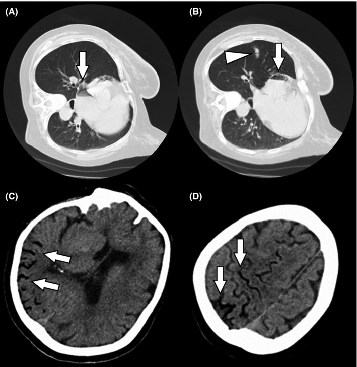
Case: A 74-year-old woman underwent computed tomography-guided transthoracic needle biopsy of a small lung mass. Immediately after the procedure, she lost consciousness. After resuscitation, her brain computed tomography scan confirmed a cerebral air embolism.
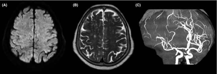
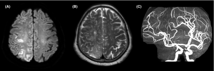
Outcome: As hyperbaric oxygenation was unavailable, she received controlled normothermia for neuroprotection. No cerebral symptoms were observed following treatment.
Conclusion: Air embolisms are rare, but fatal, complications of computed tomography-guided transthoracic needle biopsy. Therefore, clinicians should be familiar with early diagnosis and prompt treatment. Preventing hyperthermia might be effective for treating hypoxic brain injury caused by cerebral air embolisms.
Keywords: Air embolism; complication; hypoxic brain damage; ischemia; targeted temperature management.
Figures
Similar articles
-
Nonfatal systemic air embolism complicating percutaneous CT-guided transthoracic needle biopsy: four cases from a single institution.Chest. 2007 Aug;132(2):684-90. doi: 10.1378/chest.06-3030. Chest. 2007. PMID: 17699141
-
Cerebral air embolism complicating CT-guided trans-thoracic needle biopsy of the lung.Clin Respir J. 2011 Apr;5(2):e1-3. doi: 10.1111/j.1752-699X.2010.00229.x. Epub 2010 Dec 1. Clin Respir J. 2011. PMID: 21410896
-
Systemic air embolism as a complication of percutaneous computed tomography guided transthoracic lung biopsy.Ann R Coll Surg Engl. 2017 Jul;99(6):e174-e176. doi: 10.1308/rcsann.2017.0091. Ann R Coll Surg Engl. 2017. PMID: 28660818 Free PMC article.
-
Systemic air embolism following computed-tomography-guided transthoracic needle biopsy of lung lesion - a systematic search of case reports and case series.Acta Radiol Open. 2022 Jun 25;11(6):20584601221096680. doi: 10.1177/20584601221096680. eCollection 2022 Jun. Acta Radiol Open. 2022. PMID: 35770135 Free PMC article. Review.
-
Complications of CT-guided transthoracic lung biopsy : A short report on current literature and a case of systemic air embolism.Wien Klin Wochenschr. 2018 Apr;130(7-8):288-292. doi: 10.1007/s00508-018-1317-0. Epub 2018 Jan 23. Wien Klin Wochenschr. 2018. PMID: 29362884 Free PMC article. Review.
Cited by
-
Incidence, risk factors, and prognostic indicators of symptomatic air embolism after percutaneous transthoracic lung biopsy: a systematic review and pooled analysis.Eur Radiol. 2021 Apr;31(4):2022-2033. doi: 10.1007/s00330-020-07372-w. Epub 2020 Oct 13. Eur Radiol. 2021. PMID: 33051730 Free PMC article.
-
Risk factors for air embolism following computed tomography-guided percutaneous transthoracic needle biopsy: a systematic review and meta-analysis.Diagn Interv Radiol. 2023 May 31;29(3):478-491. doi: 10.4274/dir.2022.221187. Epub 2023 Mar 20. Diagn Interv Radiol. 2023. PMID: 36994842 Free PMC article.
References
-
- Tomiyama N, Yasuhara Y, Nakajima Y et al CT‐guided needle biopsy of lung lesions: a survey of severe complication based on 9783 biopsies in Japan. Eur. J. Radiol. 2006; 59: 60–4. - PubMed
-
- Van Hulst RA, Klein J, Lachmann B. Gas embolism: pathophysiology and treatment. Clin. Physiol. Funct. Imaging 2003; 23: 237–46. - PubMed
-
- Mitchell S, Gorman D. The pathophysiology of cerebral arterial gas embolism. J. Extra Corpor. Technol. 2002; 34: 18–23. - PubMed
-
- Muth CM, Shank ES. Gas embolism. N. Engl. J. Med. 2000; 342: 476–82. - PubMed
Publication types
LinkOut - more resources
Full Text Sources
Other Literature Sources
Research Materials
